External users: registration to be carried out only through I-STEM portal
Additional information about sample and analysis details should be filled in the pdf form provided in the I-STEM portal under “DOWNLOAD CSRF”
Internal users (IITB): registration to be carried out only through DRONA portal
Additional information about sample and analysis details should be filled in the pdf form provided here.
.
Category
- Spectroscopy and Spectrometry » Optical Spectroscopy
Booking Details
Facility Management Team and Location
Facility Features, Working Principle and Specifications
Facility Description
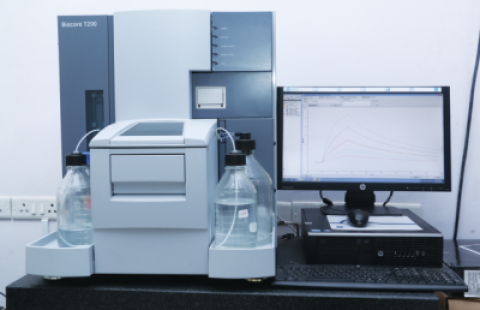
Surface Plasmon Resonance (SPR) instruments is an optical spectroscopy that allow measuring binding constants of molecular interactions. It is a label-free, real- time technique. Capable to measure binding affinities and kinetics for bio-molecular interactions. This facility caters to a wide range of applications in drug discovery, protein interaction studies, antibody validation, lipids, nucleic acids binding kinetics, drugs small molecule screening, Fab, Mab screening,epitope binning.
Surface Plasmon resonance (SPR) instrument use an optical method to measure the changes in refractive index within about 150 nm from the sensor surface. It monitors molecular interactions in real time, using a non-invasive label-free technology that responds to changes in the concentration of molecules at a sensor surface as molecules bind to or dissociate from the surface. The essential components of a Biacore analytical system are sensor chip, optical detector and integrated microfluidic cartridge (IFC) . To study the interaction between two binding partners, one partner is attached to the sensor surface (ligand) and the other is passed over the surface through flow cells in sample solution (analyte). As the analyte binds to the ligand the accumulation of protein on the sensor surface causes an changes in refractive index. A sensorgram is a plot of response against time, showing the progress of the interaction.The SPR response is directly proportional to the change in mass concentration close to the sensor chip surface.
- Detection technology: Surface Plasmon Resonance (SPR) biosensor
- Information provided: Kinetic and affinity data (KD, ka, and kd), specificity, selectivity, concentration, and thermodynamic data
- Data presentation: Result tables, result plots, and real-time monitoring of sensorgrams
- Analysis time per cycle:
- Typically, 2 to 15 min
- Automation: 48 h unattended operation
- Sample type: LMW drug candidates to high molecular weight proteins (also DNA, RNA, polysaccharides, lipids, cells, and viruses) in various sample environments (e.g., in DMSO-containing buffers, plasma, and serum)
- Required sample injection volume: Plus 20 - 50 µl volume (application dependent)
- Injection volume: 2 to 350 µl
- Flow rate range: From 1 to 100 µl/min
- Flow cell volume: 0.06 µl
- Flow cell height: 40 µm
- Sample/reagent capacity: 1 x 96-or 384-well microplate and up to 33 reagent vials
- Analysis temperature: 4°C to 45°C (maximum 20°C below ambient temperature)
- Sample storage: 4°C to 45°C (maximum 15°C below ambient temperature)
- Sample refractive index range: 1.33 to 1.40
- Buffer selector: Automatic switching between 4 buffers
- In-line reference subtraction: Automatic
Sample Preparation, User Instructions and Precautionary Measures
- Experiments should be discussed with PI/TA and the facility in-charge before proceeding. As SPR involves an assay development hence many procedures need to be planned before. (like choice of buffer, sensor chips, immobilization methods, and many critical parameters)
- Purity of samples is extremely important for generating good data.
- Protein concentrations should be measured accurately before starting the experiment.
- The molecular weight as well as the pI of the proteins should be known before immobilization.
- All buffers should be filtered through 0.22 μm filters and degassed.
- Do not degas buffers containing detergent. Add detergent after degassing.
- For organic solvent containing buffer, filter using organic solvent resistant membrane.
- Cell extracts and nanoparticles can block integrated micro fluidic cartridges and syringes.
- Any query regarding your SPR experiment can be emailed on spr.bios@iitb.ac.in
- Appointments will be provided as per queue and the user will be informed about the same.
- Kindly perform some literature review on this kind of work performed on either same or similar samples. Accumulate as much information as possible for better quality results.
Charges for Analytical Services in Different Categories
External SAmples
Category |
Charges ( INR) |
IIT Bombay | 1500 |
Other Academic Institutes | 3000+ 18% GST |
National Labs |
7500+ 18% GST |
| Industries | 15000+ 18% GST |
Start-up (SINE incubation) | 7500+ 18% GST
|
Monash IIT B |
1500+ 18% GST |
| SAARCS countries & African Countries (LowIncome) – Academic | 7500 |
| SAARCS countries & African Countries (LowIncome) – Industries | 15000 |
| Other countries – Academic | 15000 |
| Other countries - Industries | 30000 |
* Additional charges: Sensor chip charge, Maintenance charge is applicable*
*Cost and experimental details for other types of assays can be discussed as per requirement, method development charges will be applicable
Applications
- Assay Development for binding studies for Biomolecule, Small molecules, Drugs binding studies.
- Binding study of these molecules in crude lysates, whole cells, etc
- Interaction Kinetics analysis of Proteins, Nucleic acid, carbohydrates, Lipids, Small molecule, Drugs.
- Mab screening.
- Epitope Binning.
- Immunogenicity testing
- Calibration Free Concentration analysis
- Sensor development for testing
Sample Details
Sample form: Purified Proteins, Peptides ,Lipids, Liposomes ,Nucleic acids, small molecule, mabs.
SOP, Lab Policies and Other Details
Publications
Publications:
Sharma, K., Mehra, S., Sawner, A. S., Markam, P. S., Panigrahi, R., Navalkar, A., Chatterjee, D., Kumar, R., Kadu, P., Patel, K., Ray, S., Kumar, A., & Maji, S. K. (2020). Effect of Disease-Associated P123H and V70M Mutations on β-Synuclein Fibrillation. ACS chemical neuroscience, 11(18), 2836–2848.
Harish, M., & Venkatraman, P. (2021). Evolution of biophysical tools for quantitative protein interactions and drug discovery. Emerging topics in life sciences, 5(1), 1–12.
Chatterjee, D., Jacob, R. S., Ray, S., Navalkar, A., Singh, N., Sengupta, S., Gadhe, L., Kadu, P., Datta, D., Paul, A., Arunima, S., Mehra, S., Pindi, C., Kumar, S., Singru, P., Senapati, S., & Maji, S. K. (2022). Co-aggregation and secondary nucleation in the life cycle of human prolactin/galanin functional amyloids. eLife, 11, e73835. Top of Form
Gaikwad, D. D., Bangar, N. S., Apte, M. M., Gvalani, A., & Tupe, R. S. (2022). Mineralocorticoid interaction with glycated albumin downregulates NRF–2 signaling pathway in renal cells: Insights into diabetic nephropathy. International Journal of Biological Macromolecules, 220, 837-851.
Kadu, P., Gadhe, L., Navalkar, A., Patel, K., Kumar, R., Sastry, M., & Maji, S. K. (2022). Charge and hydrophobicity of amyloidogenic protein/peptide templates regulate the growth and morphology of gold nanoparticles. Nanoscale, 14(40), 15021-15033.
Dey, A., Mitra, D., Rachineni, K., Khatri, L. R., Paithankar, H., Vajpai, N., & Kumar, A. (2022). Mapping of Methyl Epitopes of a Peptide‐Drug with Its Receptor by 2D STDD‐Methyl TROSY NMR Spectroscopy. ChemBioChem, e202200489.
Panigrahi, R., Krishnan, R., Singh, J. S., Padinhateeri, R., & Kumar, A. (2023). SUMO1 hinders α‐Synuclein fibrillation by inducing structural compaction. Protein Science, e4632.
Bhambid, M., Dey, V., Walunj, S., & Patankar, S. (2023). Toxoplasma Gondii Importin α Shows Weak Auto-Inhibition. The Protein Journal, 42(4), 327–342
Poudyal M, Patel K, Gadhe L, Sawner AS, Kadu P, Datta D, Mukherjee S, Ray S, Navalkar A, Maiti S, Chatterjee D, Devi J, Bera R, Gahlot N, Joseph J, Padinhateeri R, Maji SK. Intermolecular interactions underlie protein/peptide phase separation irrespective of sequence and structure at crowded milieu. Nat Commun. 2023 Oct 4;14(1):6199.
Venkatramani, A., Ashtam, A., & Panda, D. (2024). EB1 Increases the Dynamics of Tau Droplets and Inhibits Tau Aggregation: Implications in Tauopathies. ACS chemical neuroscience, 15(6), 1219–1233.
