External users: registration to be carried out only through I-STEM portal
Additional information about sample and analysis details should be filled in the pdf form provided in the I-STEM portal under “DOWNLOAD CSRF”
Internal users (IITB): registration to be carried out only through DRONA portal
Additional information about sample and analysis details should be filled in the pdf form provided here.
.
Category
- Spectroscopy and Spectrometry » Raman Spectrometer
Booking Details
Facility Management Team and Location
chinnasamys@iitb.ac.in
gmathew@iitb.ac.in
Facility Features, Working Principle and Specifications
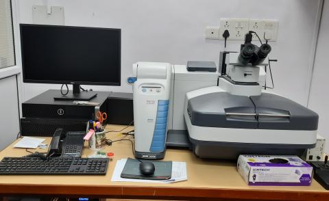
Facility Description
Laser Raman Confocal Imaging Spectroscope is based on scattering of light and gives information on chemical and structural characteristics of the sample with minimal or no sample preparation.
When a sample is irradiated with an intense monochromatic light source (usually a laser), most of the radiation is scattered by the sample at the same wavelength as that of the incoming laser radiation in a process known as Rayleigh scattering. However, a small proportion of the incoming light – approximately one photon out of a million – is scattered at a wavelength that is shifted from the original laser wavelength. This shift is called the Raman shift. The difference in energy between the incident laser excitation and the Raman scattered light is equal to a vibrational transition energy of the molecule. By analyzing the inelastically scattered Raman light one can build up a Raman spectrum. The Raman spectrum is plotted as a Raman shift relative to the excitation wavelength.
Lasers: 532 nm and 785 nm,
Spectral range: 50- 3500cm-1
Spectral Resolution: 5 cm-1,
Confocal depth resolution is down to 2 microns.
Sample Preparation, User Instructions and Precautionary Measures
- The sample’s height should be smaller than 2 cm when placed on the motorized sample stage.
- The sample’s upper surface should be relatively smooth and flat to minimize the chance of ramming objective into sample.
- Geological Samples should be polished accordingly.
- Kindly mark the sample area to be observed.
- Base of the sample should be flat.
Charges for Analytical Services in Different Categories
| Categories and Charges (in Rs.) (charges per hour) | |||||
| Type of testing/ experiment | Industry | National Labs | Universities (External) | Internal Users | Department Users |
| Raman Analysis | 2000 | 1200 | 800 | 200 | 200 |
| Imaging/ Mapping | 5000 | 3000 | 2000 | 500 | 500 |
(Charges excluding of 18% GST)
Applications
Earth sciences, pharmaceutical, polymer, battery, energy, semiconductor, microplastics, and many other materials science applications
Sample Details
User should be aware of the properties of the sample.
Sample form: Powder/Pellet/ On glass substrate/Film.
Liquid samples should be submitted in vials.
No
The bulk sample should be less than 8 mm x 8 mm and base of the sample should be flat for mounting on sample holder.