External users: registration to be carried out only through I-STEM portal
Additional information about sample and analysis details should be filled in the pdf form provided in the I-STEM portal under “DOWNLOAD CSRF”
Internal users (IITB): registration to be carried out only through DRONA portal
Additional information about sample and analysis details should be filled in the pdf form provided here.
.
Category
- Microscopy and Imaging » X-ray Microscopy
Booking Details
Facility Management Team and Location
Facility Features, Working Principle and Specifications
Facility Description
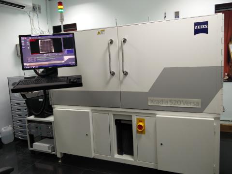
Four Dimensional X-ray Microscopy, also known as X-ray Computed Tomography (CT) is a nondestructive technique for visualizing interior features within solid objects, and for obtaining digital information on their 3-D geometries and properties.
The sample is imaged from different directions, ideally across an angular range of at least 180°. A single image at one particular angle is called a projection. Computer algorithms can be used to reconstruct the internal, 3-dimensional (3D) structure of the sample from a series of projections. The reconstructed volume can be visualized in different ways; for example slice by slice (also referred to as virtual cross-sectioning), or by rendering a 3D view of individual internal features.
Working Principle
X-rays are directed at an object from multiple orientations and the decrease in intensity along a series of linear paths is measured. This decrease is characterized by Beer's Law, which describes intensity reduction as a function of X-ray energy, path length, and material linear attenuation coefficient. A specialized algorithm is then used to reconstruct the distribution of X-ray attenuation in the volume being imaged.
Key features:
•Non-destructive interior tomography uniquely enabled by Scout-and-Zoom
•True spatial resolution of 700 nm
•Minimum achievable voxel of 70 nm
•Two-stage magnification that provides resolution at a distance (RaaD), delivering large, flexible working distances while maintaining submicron resolution
Sample Preparation, User Instructions and Precautionary Measures
1. Sample should be in solid form. No liquid or powdered samples are allowed.
2. Max sample size permitted is 3cm * 3cm * 3cm.
3. For better results, sample should be much smaller than the maximum sample size.
4. The sample should have one flat surface for the ease of mounting on the sample holder.
5.The samples should not be hazardous, chemically or physically unstable or reactive when exposed to X-rays or during handle.
- Slots will be provided on a first-come-first-served basis.
- The samples should conform to the attached requisition form and be provided along with a duly signed Non-dangerous material undertaking Form.
- Appointment: The users will be informed about their date and time-slot by email. If the day and time-slot is not suitable for you, an email request should be sent immediately for an alternate slot.
- Time frame for providing the experimental data depends on the availability of the machines and various external factors.
- The scan time is dependent on the sample type, physical dimensions, magnification, type of tomography and other factors.
- Sample Submission (for externals): Samples are to be brought in-person on the date of your appointment for analysis or it can be couriered to Prof. Asim Tewari, Convener, Central FDXM Facility, SAIF, IIT Bombay, Powai, Mumbai - 400076, along with the Request Letter and Sample Requisition form and Non-dangerous Material undertaking Form.
- Results: Since very large volume (several GBs) of experimental data is produced, the data would be provided on an external hard drive (formatted) which needs to be provided along with the samples.
- Return shipping charges need to be paid, if the samples need to be returned to the originator after testing. We are not responsible if the samples or external hard drives are damaged in transit.
- The experimental data will be provided in the form of a stack of TIFF files and no further analyses or interpretation of the data will be provided.
- The experimental data would only be stored at our facility for only 90 days after returning the hard drives.
Charges for Analytical Services in Different Categories
S.No. | Category | Charge ratio. | GST @ 18% |
1. | IITB (TAs) | 375 | No GST |
2. | IITB Students | 750 | No GST |
3. | IITB-Monash Students | 750 | + GST |
4. | Academic Institutes | 1500 | + GST |
5. | National Labs | 3750 | + GST |
6. | Sine (letter from SINE reqd.) | 3750 | + GST |
7. | Research Park (MSME) (letter from RP reqd.) | 3750 | + GST |
8. | Research Park (Big Industry partners) (letter from RP reqd.) and MSME not associated with RP (appropriate certificate required) | 5625 | + GST |
9. | Industries | 7500 | + GST |
Applications
- Materials Research
Characterize materials, observe fracture mechanics, investigate properties at multiple length scales, quantify and characterize microstructural evolution. Perform in situ and 4D (time dependent) studies to understand the impact of heating, cooling, oxidation, wetting, tension, tensile compression, imbibition, drainage and other simulated environmental studies.
- Life Sciences
Perform virtual histology, visualize cellular and subcellular features, and characterize submicron structures in samples that are inches to centimeters in size.
- Natural Resources
Characterize and quantify pore structures, measure fluid flow, study carbon sequestration processes, analyze tailings to maximize mining efforts. Accurate 3D submicron support for digital rock simulations, in situ multiphase fluid flow studies, and 3D mineralogy.
- Electronics
Optimize processes development, study package reliability, perform failure analysis, and analyze package construction. Non-destructive submicron imaging of intact packages for defect re-localization and characterization.
- Pharmaceuticals
Enables to measure the thickness and uniformity of coatings applied to tablets and capsules. Visualize the distribution of ingredients in the final dosage, characterize foreign material based on density, shape or size, detection of anomalies in formulations. High contrast tomography helps in establishing correlations between coating density, drug product performance and product expiry.
Sample Details
NA
NA
NA
NA
NA
NA
NA
SOP, Lab Policies and Other Details
Publications
Dutta, S.; Kumar, S.; Singh, H.; Khan, M.A.; Barai, A.; Tewari, A.; Rana, R.S.; Bera, S.; Sen, S.; Sahni, A. Chemical evidence of preserved collagen in 54-million-year-old fish vertebrae. Palaeontology 2020, 63, 195–202
Deepoo Kumar ;Ross Cunningham ; PC Pistorius. Use of X-Ray Microtomography to Determine Volume Fraction and 3D Morphology of Inclusion Clusters in Steel. AISTech 2018,1493-1500.
Bankim Mahanta, Vikram Vishal, Nikhil Sirdesai, PG Ranjith, TN Singh,
Progressive deformation and pore network attributes of sandstone at in-situ stress states using computed tomography. Engineering Fracture Mechanics,Volume 252, 2021, 107833,ISSN 0013-7944.
Vikram Vishal, Debanjan Chandra. Mechanical response and strain localization in coal under uniaxial loading, using digital volume correlation on X-ray tomography images.International Journal of Rock Mechanics and Mining Sciences, Volume 154,
2022, 105103,ISSN 1365-1609.
Rathi, P.; Jha, M. K.; Hata, K.; Subramaniam, C. Real-Time, Wearable, Biomechanical Movement Capture of Both Humans and Robots with Metal-Free Electrodes. ACS Omega 2017, 2, 4132– 4142, DOI: 10.1021/acsomega.7b00491
