.
Category
- Microscopy and Imaging » Electron Microscopy
Booking Details
Facility Management Team and Location
Facility Features, Working Principle and Specifications
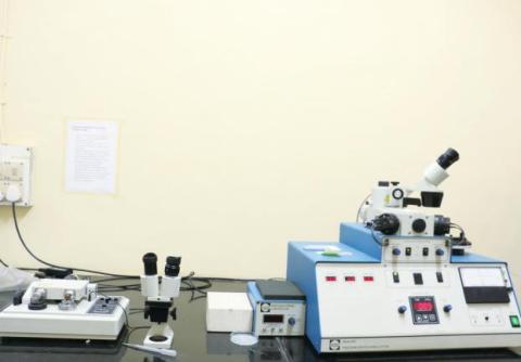
TEM Sample Preparation
The quality of Transmission Electron Microscopy (TEM) images heavily relies on the preparation of the sample. This process includes:
Thinning the sample through cutting.
Grinding to achieve the required thickness.
Dimpling the sample surface.
Ion-milling to further reduce thickness and improve transparency to electrons.
It's crucial to ensure that these steps do not alter the intrinsic properties of the material being studied.
Variable Low Speed Diamond Saw
Materials (ceramics and metals) can be reduced to 0.5mm in thickness using this technique, which uses a diamond blade, with the wheel speed varying between 100 to 400 rpm.Tuned Piezo Cutting Tool
This is used for cutting the material (both ceramic and metal) in the desired size and shape, so that it can be placed inside the TEM sample holder
Ratings and Specifications
Disc Cutter External PSU (Power Supply Unit)
Input Voltage: 100-240 VAC (~)
Output Voltage: 24VDC
Output Power: 30W
Frequency: 47-63 Hz
Mains supply voltage fluctuations are not to exceed 10% of the nominal supply voltage
Disc Cutter Power Jack ("DC in")
DC Input Voltage: 24 VDC
DC Input Current: 1.25 A
Stereo Microscope Power Jack ("DC Out")
Used to power optional microscope
Output Voltage: 24 VDC
Output Current: 0.4A
Environmental Conditions
Non-operating relative humidity (non-condensing): 10-90%
Storage temperature range: 0-65°C
Operating Humidity Range: 20-80%
Operating maximum thermal gradient: 15°C/hr
Operating temperature range: 16-24°C
(Information partly obtained from GATAN Manual)Disc Punch
Used only for metallic sheets. The Disc punch prepares specimen discs of 3 mm in diameter and material thicknesses ranging from 10 µm to above 100 µm.Specimen Mounting Hot Plate
This is used for firmly attaching specimen discs to stubs during grinding. This is done by using a low melting point wax polymer, heated on the hot plate, to form a strong, thin, hard adhesive bond. Operating temperature range is up to 130°C.Disc Grinder
This is used for holding the stub with the specimen attached for grinding down to a certain thickness. The GATAN Model 623 Disc Grinder possesses superior resolution and preloading of the specimen drive screw to result in excellent control of specimen thickness. By using the weight of the grinder it limits the maximum polishing pressure the operator is able to reduce specimen damage to a minimum. The samples are polished using different grit silicon carbide papers such as 40 µm, 15 µm, and 5 µm, in sequence.(Information partly obtained from GATAN Manual)Dimple Grinder
After manual grinding, using a disc grinder, precision thinning near the central region of the 3 mm disc specimen (or forming a dimple) is performed using a dimple grinder.
Specifications:
External PSU Input Voltage: 100-240 VAC (~)
Output Voltage: 24 VDC
Output Power: 30W
Frequency: 47-63 Hz
Power Jack ("DC in")
DC Input Voltage: 24 VDC
DC Input Current: 1.25A
Stereo Microscope Power Jack ("DC out")
Output Voltage: 24 VDC
Output Current: 0.4A
Environmental Conditions
Non-operating relative humidity: 10-90%
Storage temperature range: 0-65°C
Operating humidity range: 20-80%
Operating maximum thermal gradient: 15°C/hr
Operating temperature range: 16-24°CPrecision Ion Polishing System (PIPS)
After dimple grinding, the final stage of precision polishing (thinning) of the 'dimpled' region up to electron transparency is done with the PIPS.
Specifications:
Input Voltage: 100/120/240V
Input Current: 3/3/1.5A
Frequency: 50/60Hz
Output Voltage: 120VAC 50/60Hz
Output Current: 3.0 A Max.
Non-operating relative humidity (non-condensing): 10-90%
Storage temperature range: 0-65°C
Operating humidity range: 20-80%
Operating maximum thermal gradient: 15°C/hr
Operating temperature range: 5-35°C
For indoor use only
(Information partly obtained from GATAN Manual)
Instructions for Registration, Sample Preparation, User Instructions and Precautionary Measures
TEM Sample Preparation Guidelines
Maximum of two samples per slot, scheduled either in the forenoon or afternoon on any working day. Note that preparation time may exceed slot duration.
Sample dimensions must be adequate for TEM preparation processes.
The user must be present and available during the entire TEM sample preparation process.
We shall accept online registration only through the IRCC webpage. If you need to cancel your slot, send an email immediately to with an explanation.
Slots will be provided on a first-come-first-served basis.
Users must be present during the entire slot.
Charges for Analytical Services in Different Categories
Applications
TEM Sample Preparation Facility
The Transmission Electron Microscopy (TEM) sample preparation facility specializes in creating thin samples that are electron-transparent for TEM analysis. Equipped with advanced tools and techniques, the facility can produce both planar and cross-sectional samples across a wide range of material types.
Methods employed include focused ion beam (FIB) milling, mechanical polishing, and other precise techniques to ensure the samples meet the stringent requirements for TEM analysis.
This capability is crucial for research in materials science, nanotechnology, and other fields where high-resolution imaging of material structures at the atomic level is essential.
