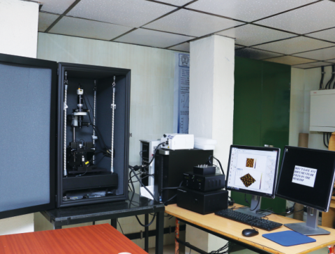
.
Category
- Microscopy and Imaging » Force Microscopy
Booking Details
Facility Management Team and Location
Facility Features, Working Principle and Specifications
Scanners : Lateral range X-Y imaging area up to 50 μm x 50 μm, Vertical (Z) range: 7μm;
AFM stands for Atomic Force Microscopy or Atomic Force Microscope and is often called the "Eye of Nanotechnology". AFM, also referred to as SPM or Scanning Probe Microscopy, is a high-resolution imaging technique that can resolve features as small as an atomic lattice in the real space. It allows researchers to observe and manipulate molecular and atomic level features.
AFM works by bringing a cantilever tip in contact with the surface to be imaged. An ionic repulsive force from the surface applied to the tip bends the cantilever upwards. The amount of bending, measured by a laser spot reflected on to a split photo detector, can be used to calculate the force. By keeping the force constant while scanning the tip across the surface, the vertical movement of the tip follows the surface profile and is recorded as the surface topography by the AFM.
The predecessor of AFM is STM, Scanning Tunneling Microscopy or the Scanning Tunneling Microscope, was invented in 1981 by G. Binnig and H. Rohrer who shared the 1986 Nobel Prize in Physics for their invention. An excellent technique, STM is limited to imaging conducting surfaces.
Atomic Force Microscopy has much broader potential and application because it can be used for imaging any conducting or non-conducting surface. The number of applications for AFM has exploded since it was invented in 1986 and now encompass many fields of nanoscience and nanotechnology. It provides the ability to view and understand events as they occur at the molecular level which will increase our understanding of how systems work and lead to new discoveries in many fields. These include life science, materials science, electrochemistry, polymer science, biophysics, nanotechnology, and biotechnology.
Instructions for Registration, Sample Preparation, User Instructions and Precautionary Measures
Registration: To avail the SPM-I@MEMS (Atomic Force Microscope) Facility, located in the Room
Number G021, Ground floor of the Department of Metallurgical Engineering and Materials Science, IIT
Bombay, registration is absolutely essential.
Registration Process:
I) Internal Users:
User within IIT Bombay can apply from
https://drona.ircc.iitb.ac.in/ircc/NewFac/CentralFacilityIndex.jsp?facilityCode=MT005
The form should be completely filled up and all the sample details must be provided in the requisition
form. The user needs to stay in the laboratory during the entire duration of the experiment, as per the slot
assigned. If a user wishes to change his/her time slot, an email should be sent immediately to
spm_mems@iitb.ac.in requesting change in appointment.
II) External Users:
Registration Link:
https://rnd.iitb.ac.in/research-facility/scanning-probe-microscope-facility-i
External Users can register through above link. External Payment should be made in advance by a
Demand Draft (DD) drawn in favour of “The Registrar, IIT Bombay, P and C Account". The same should
be sent to “Prof. Tanushree H. Choudhury, Convener, Scanning Probe Microscope Facility (SPM) -I, Central Facility,
Department of Metallurgical Engineering and Materials Science, IIT Bombay, Powai, Mumbai 400076”.
Appointments will be given after complete registration and payment. Any query related to the sample
preparation should be emailed to “spm_mems@iitb.ac.in”
Academic Institutions:
You can come personally or send a letter from the Guide/HoD on the Institution’s Original Letter Head
stating that the analysis is for research purpose, to qualify for academic concession along with the
Registration Form and Demand draft. The letter should be addressed to “Prof. Tanushree H. Choudhury, Convener,
Scanning Probe Microscope Facility (SPM) -I, Central Facility, Department of Metallurgical Engineering and Materials Science, IIT Bombay, Powai, Mumbai 400076”
Industry & Non- Government Agencies:
You can come personally or send a letter signed by an authorized signatory of your Institution on Original
Letter Head along with the Registration Form and Demand draft. The letter should be addressed to “Prof. Tanushree H. Choudhury, Convener, Scanning Probe Microscope Facility (SPM) -I, Central Facility, Department of
Metallurgical Engineering and Materials Science, IIT Bombay, Powai, Mumbai 400076”
National R & D Lab’s:
You can come personally or send a letter signed by an authorized signatory of your Institution on Original
Letter Head stating that the analysis is for research purpose along with the Registration Form and Demand
draft. The letter should be addressed to “Prof. Tanushree H. Choudhury, Convener, Scanning Probe Microscope
Facility (SPM) -I, Central Facility, Department of Metallurgical Engineering and Materials Science, IIT
Bombay, Powai, Mumbai 400076”
You are requested to check the appropriate box in the registration form if you agree (or not) to
acknowledge the Scanning Probe Microscope Facility -I (SPM) @ MEMS of IIT BOMBAY in your
Publications/Reports/Thesis/Product development/prototype development/ proof of concept in which
the data is used.
If you agree, you are requested to mention in your request letter that “We agree to acknowledge the
Scanning Probe Microscope -I (SPM) Central Facility of IIT Bombay when the data obtained from this
facility are used in our Publications/Reports/Thesis/Product development/prototype development/ proof
of concept”. List of such acknowledgements with appropriate reference will be communicated to SPM-I
@MEMS facility vide email to “spm_mems@iitb.ac.in”.
Kindly send the publication reference (Journal name/volume number/names of the authors/date of issue
of the publication etc) to us as the continuing functioning of the lab needs this feedback.
1. The experimental data provided is only for research / development purposes. These cannot
be used as certificates in legal disputes.
2. The users should know the approximate roughness of the sample before submission for
analysis.
3. MSDS (Material Safety Data Sheet) should be given along with samples to ensure that the
samples are not toxic or hazardous. Samples will not be accepted unless accompanied by
MSDS.
4. The Sample will be mounted on steel disc with the help of double-sided adhesive tape for
strong adhesion, so the sample may get damaged during removal.
5. Please bring a CD with you for data collection.
Charges for Analytical Services in Different Categories
Applications
Life Science
Material Science
Polymer Science
Electrical characterization
Nanolithography
Nano-grafting
Biotechnology
Sample Details
SOP, Lab Policies and Other Details
Publications
S. Sarkar, D. Majhi, S. Kurup, Dipti Gupta*, “Photonic Cured Metal Oxides for Low-Cost High-Performance Low Voltage Flexible and Transparent Thin Film Transistors”, ACS Applied Electronic Materials, v4, p2442, (2022)
Javed Alam Khan, Ajay Singh Panwar, Dipti Gupta, “Domain modulation and energetic disorder in ternary bulk-heterojunction organic solar cells”, Organic Electronics, v102, p106376, (2022) (I.F.)
J Khan, Ramakant Sharma, Ajay Singh Panwar, Dipti Gupta, “Impact of non-fullerene acceptors and solvent additive on the nanomorphology, device performance, and photostability of PTB7-Th polymer based organic solar cells”, Journal of Physics D: Applied Physics, v55, p495503
