External users: registration to be carried out only through I-STEM portal
Additional information about sample and analysis details should be filled in the pdf form provided in the I-STEM portal under “DOWNLOAD CSRF”
Internal users (IITB): registration to be carried out only through DRONA portal
Additional information about sample and analysis details should be filled in the pdf form provided here.
.
Category
- Diffraction » Single Crystal XRD: Protein
Booking Details
Facility Management Team and Location
Facility Features, Working Principle and Specifications
Facility Description
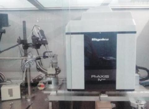
Our protein crystallography facility is a dedicated laboratory designed to facilitate the research and analysis of protein structures through X-ray crystallography, a leading and widely used method for determining the 3D atomic-level structures of proteins. The facility is equipped with an X-ray diffractometer and automated crystallization robots to support efficient and high-quality experimentation.
Protein X-ray crystallography is a powerful technique used to determine the 3D atomic structure of proteins. The process involves several key steps:
- Crystallisation: Purified protein is crystallised into highly ordered, repeating lattices that allow for X-ray diffraction.
- X-ray Exposure: The protein crystal is exposed to X-rays, which are scattered by the atoms in the crystal, producing a diffraction pattern.
- Data Collection: A series of diffraction patterns is collected by rotating the crystal in the X-ray beam, capturing data from different angles.
- Phase Problem: The diffraction data provide amplitude and orientation, but not phase information. Phasing methods like molecular replacement or heavy atom derivatization are used to solve this problem.
- Electron Density Map: The data, with phase information, are used to calculate an electron density map, which shows the positions of atoms in the crystal.
- Model Building and Refinement: Researchers build an atomic model based on the electron density map and refine it by adjusting atom positions to minimise discrepancies with the data.
- Validation: The final model is validated for accuracy using various quality metrics and tests.
Diffractometer:
- Maximum Rated Output: 1.2 kW
- Rated Tube Voltage-Current: 40 kV; 30 mA
- Target: Copper (Cu)
- Radiation Enclosure: Full safety shielding with plastic cover
- Scanning Mode: 0-180° ω scan, 0-180° θ scan
- Optics: Multilayer confocal type
- Detector: Rigaku R Axis IV++
- Beam Size at the Sample: 100 µm
Mosquito Crystallization Robot:
- Designed for screening soluble and membrane proteins (LCP).
- Capable of performing hanging drop, sitting drop, microbatch, microseeding, and additive screening.
- Automated syringe dispenser for accurate liquid handling.
- Highly precise and rapid automated plate setups.
- Dispense volume range: 25 nL to 1200 nL (protein), 70 µL (well solution).
- Ensures zero cross-contamination with no washing steps required.
Phoenix Crystallization Robot:
- Ideal for screening soluble proteins.
- Supports sitting drop, hanging drop, and microbatch reactions.
- Dispense volume range: 100 nL to 100 µL.
- Rapid dispensing (< 50 seconds) of screen and protein, minimizing evaporation issues.
- Flexible dispensing needles.
- Multiple proteins can be quickly dispensed into multi-well plates.
Sample Preparation, User Instructions and Precautionary Measures
- Well-isolated protein crystals from crystallisation plates should be supplied for the slot.
- Crystals can also be flash-frozen in liquid nitrogen with the appropriate cryoprotectant, stored in a Dewar, and sent to us for the slot.
- Users must provide their own pre-prepared cryoprotectant solutions.
- Cover-slips, soaking bridges, and loops suitable for your proteins will be provided upon request, with additional charges applicable.
- For crystallisation, purified proteins with a sufficient concentration should be provided.
- The user must be present during the X-ray diffraction of the crystal or setting up crystallisation trays.
- Diffraction images will be provided on a CD. Please bring your own CD, as USB drives are strictly prohibited due to virus concerns.
- Samples and measurement data must be collected immediately after the diffraction is completed, with a maximum allowed duration of one week.
Charges for Analytical Services in Different Categories
Charges for Internal Users (18% GST applicable)
| Sr. No. | Organization | Screening per day | Data collection per sample | Crystallization tray setup using robots per tray |
| 1 | Academic Institution | 3,500/- | 5,000/- | 3000/- |
| 2 | Industries & Non – Government Agencies | 20,000/- | 50,000/- | 25,000/- |
Charges for External Users (18% GST applicable)
| Sr. No. | Organization | Screening per day | Data collection per sample | Crystallization tray setup using robots per tray |
| 1 | Academic Institution | 4,500/- | 7,000/- | 4,000/- |
| 2 | Industries & Non – Government Agencies | 20,000/- | 50,000/- | 25,000/- |
Applications
- Understanding Protein Function: It reveals how protein structures relate to their biological roles, aiding in the study of enzyme mechanisms, signal transduction, and receptor-ligand interactions.
- Drug Design: Structural insights guide the development of targeted drugs, such as enzyme inhibitors, antibodies, and small molecule therapeutics.
- Protein-Ligand Interactions: Crystallography helps identify binding sites for small molecules, improving drug discovery and design.
- Studying Protein Complexes: It provides insights into how proteins interact in larger complexes, crucial for understanding cellular processes.
- Disease Mechanisms: It aids in understanding how mutations affect protein function, helping in the development of treatments for genetic diseases.
- Structural Genomics: It contributes to mapping the structures of proteins across various organisms, advancing biological knowledge.
- Viral and Bacterial Pathogens: Crystallography is used to design antiviral drugs and antibiotics by studying viral and bacterial proteins.
- Biotechnology: It supports enzyme engineering and the development of industrial enzymes and biocatalysts.
Sample Details
SOP, Lab Policies and Other Details
Publications
2022:
- Singh, J., Sahil, M., Ray, S., Dcosta, C., Panjikar, S., Krishnamoorthy, G., Mondal, J., Anand, R. "Phenol Sensing in Nature Is Modulated via a Conformational Switch Governed by Dynamic Allostery." Journal of Biological Chemistry, 2022, 298(10), 102399. DOI
- Sharma, N., Singh, S., Tanwar, A. S., Mondal, J., Anand, R. "Mechanism of Coordinated Gating and Signal Transduction in Purine Biosynthetic Enzyme Formylglycinamidine Synthetase." ACS Catalysis, 2022, 12(3), 1930–1944. DOI
- Kesari, P., Deshmukh, A., Pahelkar, N., Suryawanshi, A. B., Rathore, I., Mishra, V., Dupuis, J. H., Xiao, H., Gustchina, A., Abendroth, J., Labaied, M., Yada, R. Y., Wlodawer, A., Edwards, T. E., Lorimer, D. D., Bhaumik, P. "Structures of Plasmepsin X from Plasmodium falciparum Reveal a Novel Inactivation Mechanism of the Zymogen and Molecular Basis for Binding of Inhibitors in Mature Enzyme." Protein Science, 2022, 31(4), 882–899. DOI
- Sakunthala, A., Datta, D., Navalkar, A., Gadhe, L., Kadu, P., Patel, K., Mehra, S., Kumar, R., Chatterjee, D., Devi, J., Sengupta, K., Padinhateeri, R., Maji, S. K. "Direct Demonstration of Seed Size-Dependent α-Synuclein Amyloid Amplification." The Journal of Physical Chemistry Letters, 2022, 13(28), 6427–6438. DOI
- Kadu, P., Gadhe, L., Navalkar, A., Patel, K., Kumar, R., Sastry, M., Maji, S. K. "Charge and Hydrophobicity of Amyloidogenic Protein/Peptide Templates Regulate the Growth and Morphology of Gold Nanoparticles." Nanoscale, 2022, 14(40), 15021–15033. DOI
- Mehra, S., Ahlawat, S., Kumar, H., Datta, D., Navalkar, A., Singh, N., Patel, K., Gadhe, L., Kadu, P., Kumar, R., Jha, N. N., Sakunthala, A., Sawner, A. S., Padinhateeri, R., Udgaonkar, J. B., Agarwal, V., Maji, S. K. "α-Synuclein Aggregation Intermediates Form Fibril Polymorphs with Distinct Prion-like Properties." Journal of Molecular Biology, 2022, 434(19), 167761. DOI
- Chatterjee, D., Jacob, R. S., Ray, S., Navalkar, A., Singh, N., Sengupta, S., Gadhe, L., Kadu, P., Datta, D., Paul, A., Arunima, S., Mehra, S., Pindi, C., Kumar, S., Singru, P., Senapati, S., Maji, S. K. "Co-Aggregation and Secondary Nucleation in the Life Cycle of Human Prolactin/Galanin Functional Amyloids." eLife, 2022, 11. DOI
2021:
- Godsora, B. K. J., Prakash, P., Punekar, N. S., Bhaumik, P. "Molecular Insights into the Inhibition of Glutamate Dehydrogenase by the Dicarboxylic Acid Metabolites." Proteins: Structure, Function, and Bioinformatics, 2021, 90(3), 810–823. DOI
2020:
- Sharma, N., Ahalawat, N., Sandhu, P., Strauss, E., Mondal, J., Anand, R. "Role of Allosteric Switches and Adaptor Domains in Long-Distance Cross-Talk and Transient Tunnel Formation." Science Advances, 2020, 6(14). DOI
- Rathore, I., Mishra, V., Patel, C., Xiao, H., Gustchina, A., Wlodawer, A., Yada, R. Y., Bhaumik, P. "Activation Mechanism of Plasmepsins, Pepsin‐like Aspartic Proteases from Plasmodium, Follows a Unique Trans‐Activation Pathway." The FEBS Journal, 2020, 288(2), 678–698. DOI
- Badgujar, D. C., Anil, A., Green, A. E., Surve, M. V., Madhavan, S., Beckett, A., Prior, I. A., Godsora, B. K., Patil, S. B., More, P. K., Sarkar, S. G., Mitchell, A., Banerjee, R., Phale, P. S., Mitchell, T. J., Neill, D. R., Bhaumik, P., Banerjee, A. "Structural Insights into Loss of Function of a Pore Forming Toxin and Its Role in Pneumococcal Adaptation to an Intracellular Lifestyle." PLOS Pathogens, 2020, 16(11), e1009016. DOI
- Kadu, P., Pandey, S., Neekhra, S., Kumar, R., Gadhe, L., Srivastava, R., Sastry, M., Maji, S. K. "Machine-Free Polymerase Chain Reaction with Triangular Gold and Silver Nanoparticles." The Journal of Physical Chemistry Letters, 2020, 11(24), 10489–10496. DOI
2019:
- Yarramala, D. S., Prakash, P., Ranade, D. S., Doshi, S., Kulkarni, P. P., Bhaumik, P., Rao, C. P. "Cytotoxicity of Apo Bovine α-Lactalbumin Complexed with La3+ on Cancer Cells Supported by Its High Resolution Crystal Structure." Scientific Reports, 2019, 9(1), 1780. DOI
2018:
- Pandey, S., Phale, P. S., Bhaumik, P. "Structural Modulation of a Periplasmic Sugar-Binding Protein Probes into Its Evolutionary Ancestry." Journal of Structural Biology, 2018, 204(3), 498–506. DOI
- Mishra, V., Rathore, I., Arekar, A., Sthanam, L. K., Xiao, H., Kiso, Y., Sen, S., Patankar, S., Gustchina, A., Hidaka, K., Wlodawer, A., Yada, R. Y., Bhaumik, P. "Deciphering the Mechanism of Potent Peptidomimetic Inhibitors Targeting Plasmepsins – Biochemical and Structural Insights." The FEBS Journal, 2018, 285(16), 3077–3096. DOI
- Wangchuk, J., Prakash, P., Bhaumik, P., Kondabagil, K. "Bacteriophage N4 Large Terminase: Expression, Purification and X-Ray Crystallographic Analysis." Acta Crystallographica Section F: Structural Biology Communications, 2018, 74(4), 198–204. DOI
- Prakash, P., Punekar, N., Bhaumik, P. "Structural Basis for the Catalytic Mechanism and α-Ketoglutarate Cooperativity of Glutamate Dehydrogenase." Journal of Biological Chemistry, 2018, 293(17), 6241-6258. DOI
- Kirti, S., Patel, K., Das, S., Shrimali, P., Samanta, S., Kumar, R., Chatterjee, D., Ghosh, D., Kumar, A., Tayalia, P., Maji, S. K. "Amyloid Fibrils with Positive Charge Enhance Retroviral Transduction in Mammalian Cells." ACS Biomaterials Science & Engineering, 2018, 5(1), 126–138. DOI
- Mehra, S., Ghosh, D., Kumar, R., Mondal, M., Gadhe, L. G., Das, S., Anoop, A., Jha, N. N., Jacob, R. S., Chatterjee, D., Ray, S., Singh, N., Kumar, A., Maji, S. K. "Glycosaminoglycans Have Variable Effects on α-Synuclein Aggregation and Differentially Affect the Activities of the Resulting Amyloid Fibrils." Journal of Biological Chemistry, 2018, 293(34), 12975–12991. DOI
- Sharma, H., John, K., Gaddam, A., Navalkar, A., Maji, S. K., Agrawal, A. "A Magnet-Actuated Biomimetic Device for Isolating Biological Entities in Microwells." Scientific Reports, 2018, 8(1). DOI
2017:
- Gaded, V., Anand, R. "Selective Deamination of Mutagens by a Mycobacterial Enzyme." Journal of the American Chemical Society, 2017, 139(31), 10762–10768. DOI
- Ray, S., Maitra, A., Biswas, A., Panjikar, S., Mondal, J., Anand, R. "Functional Insights into the Mode of DNA and Ligand Binding of the Tetr Family Regulator Tylp from Streptomyces fradiae." Journal of Biological Chemistry, 2017, 292(37), 15301-15311. DOI
- Ghosh, S., Salot, S., Sengupta, S., Navalkar, A., Ghosh, D., Jacob, R., Das, S., Kumar, R., Jha, N. N., Sahay, S., Mehra, S., Mohite, G. M., Ghosh, S. K., Kombrabail, M., Krishnamoorthy, G., Chaudhari, P., Maji, S. K. "P53 Amyloid Formation Leading to Its Loss of Function: Implications in Cancer Pathogenesis." Cell Death & Differentiation, 2017, 24(10), 1784–1798. DOI
2016:
- Pandey, S., Modak, A., Phale, P. S., Bhaumik, P. "High Resolution Structures of Periplasmic Glucose-Binding Protein of Pseudomonas putida CSV86 Reveal Structural Basis of Its Substrate Specificity." Journal of Biological Chemistry, 2016, 291(15), 7844–7857. DOI
- Jacob, R. S., Das, S., Ghosh, S., Anoop, A., Jha, N. N., Khan, T., Singru, P., Kumar, A., Maji, S. K. "Amyloid Formation of Growth Hormone in Presence of Zinc: Relevance to Its Storage in Secretory Granules." Scientific Reports, 2016, 6(1). DOI
- Jacob, R. S., George, E., Singh, P. K., Salot, S., Anoop, A., Jha, N. N., Sen, S., Maji, S. K. "Cell Adhesion on Amyloid Fibrils Lacking Integrin Recognition Motif." Journal of Biological Chemistry, 2016, 291(10), 5278–5298. DOI
