External users: registration to be carried out only through I-STEM portal
Additional information about sample and analysis details should be filled in the pdf form provided in the I-STEM portal under “DOWNLOAD CSRF”
Internal users (IITB): registration to be carried out only through DRONA portal
Additional information about sample and analysis details should be filled in the pdf form provided here.
.
Category
- Microscopy and Imaging » Confocal Microscopy
Booking Details
Facility Management Team and Location
Facility Features, Working Principle and Specifications
Facility Description
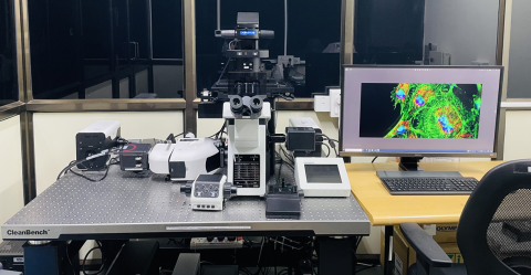
The Laser-free Confocal Microscopy Facility offers a high-speed, multi-channel imaging technique with significantly lower photobleaching compared to a conventional Point Scanning Laser Confocal Microscope.
This is a compact and fast confocal system that uses Aurox’s patented structured spinning disc and illumination technology to achieve high-resolution and high-quality confocal images.
- The resolution is approximately 300nm x 300nm in the X-Y plane and 0.6μm in the Z direction.
- The laser-free confocal unit is coupled to an IX83 Olympus microscope.
- On-stage incubation system with temperature and CO2 control for long-time live cell imaging.
- Aurox spinning disk utilizes a grid pattern instead of pinholes for optical sectioning, achieving over 50% light efficiency with the speed of a spinning disk.
- Low photo-bleaching
- Advanced image analysis software
- Full 5D confocal imaging control.
- Imaging across the scales with Clarity - 1000 microns to 0.2 microns
High compact and easy to use.
Working Principle:-
The unique optical system inside the Clarity LFC allows capturing of the images both transmitted (T) and reflected (R) by the disc to easily differentiate between in-focus+ out of Focus and out-of-focus information. Computer subtraction of these images (T–R) creates a sectioned image whereby all out-of-focus blur is effectively suppressed and only the sharp in-focus image of the sample is retained.
Using a spinning disk as the modulator/demodulator virtually eliminates residual imaging artifacts. This structured illumination pattern is used to both modulate the illumination field and demodulate the light emerging from the sample.
Aurox Confocal mode:
•Aurox patented structured illumination spinning disk
•NanoSRRF (Super Resolution Radial Fluctuations) Deconvolution
•Hamamtsu Fusion BT
•High end Xeon W-2245 core workstation
•sCMOS Hamamatsu Flash 4 V3 (95% QE) with Camera Link
•Cool LED pE 800 Light Source
Fluorescence microscope mode:
•Olympus IX83 with motorized X-Y-Z and Magnification of
10X /0.3NA (air), 40X /0.60 NA (air), 60X /1.42NA (oil), 100X /1.45NA(oil)
Sample Preparation, User Instructions and Precautionary Measures
1. Seal the fixed samples between the glass slide and the cover slip.
2. When applying for a slot, please specify the fluorophores used in your sample, including their excitation and emission spectra.
3. Live samples should be in glass coverslip-bottom Petri dishes or 6, 12, 24, and 48 well plates.
4. Please use imaging Petri dishes with coverslip bottoms if oil immersion objectives are to be used.
Instructions for users: -
- Standard slot durations are for 2 hours for fixed samples.
- USB drives are strictly prohibited to minimize virus-related issues. The data can be shared in the cloud. All data must be transferred within 7 days of imaging. Without exception.
Charges for Analytical Services in Different Categories
Charges for Internal and External Users of Laser-free Confocal Microscopy Facility@ BSBE
1 slot = 2hrs
Category | User Charges (INR) per slot* |
IITB (TA’s) | 200 |
IIT Bombay Students | 400 |
IITB-Monash Students | 400 + GST |
Other Academic Institutes | 800 + GST |
National Labs | 2000 + GST |
Sine (letter from SINE reqd.) | 2000 + GST |
Research Park (MSME) (letter from RP reqd.) | 2000 + GST |
Research Park (Big Industry partners) (letter from RP reqd.) and MSME not associated with RP (appropriate certificate required)
| 3000 + GST |
Industrial User | 4000 + GST |
Applications
- Live cell time lapse imaging
- Fast dynamic imaging
- Fixed cell imaging
- Developmental Biology
- Plant Biology
- Stem cell research
- Bioluminescent proteins
Sample Details
CO2
SOP, Lab Policies and Other Details
Publications
NA
