External users: registration to be carried out only through I-STEM portal
Additional information about sample and analysis details should be filled in the pdf form provided in the I-STEM portal under “DOWNLOAD CSRF”
Internal users (IITB): registration to be carried out only through DRONA portal
Additional information about sample and analysis details should be filled in the pdf form provided here.
.
Category
- Microscopy and Imaging » Confocal Microscopy
Booking Details
Facility Management Team and Location
Facility Features, Working Principle and Specifications
Facility Description
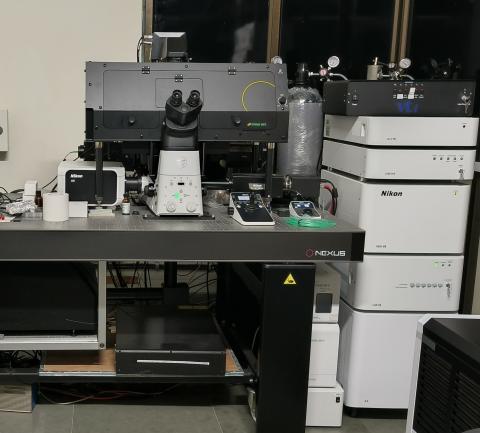
Super-resolution microscopy is a relatively new method compared to confocal microscopy, and ongoing developments aim to improve its standards. While confocal microscopy has been praised for its high contrast, recent methods show that super-resolution microscopy, specifically via the instant-structured illumination microscopy (i-SIM) technique, is capable of capturing reflectance imaging with more than double the resolution of confocal microscopy. This increased resolving capability has been applied to imaging nanoparticle uptake and localization inside cells. These developments contribute to super-resolution microscopy becoming a validated alternative to confocal microscopy for imaging subcellular activity. Whilst maintaining the necessary temporal resolution and at a low photo bleaching level, VT-iSIM offers a spatial resolution of 100nm and axial resolution of 300nm.
Super-resolution microscopy allows us to see the smallest structures in living cells that are not visible with standard widefield or confocal fluorescence microscopy. This Stochastic Optical Reconstruction Microscopy (N-STORM) technique provides a 3D resolution that is much higher than the diffraction limit, giving us new insights into the organization of cells and the dynamics of bio-molecular assemblies at a near-molecular level. Super-resolution microscopy overcomes the diffraction barrier, enabling "nanoscopy" with greatly improved optical resolution down to 20nm. This method relies on the physical or chemical properties of nearby fluorophores to distinguish them from each other.
VT-iSIM Structured Illumination Microscopy:
The leading ISM technique for live cell imaging is Instant SIM and in particular, the VT-iSIM, which allows for super-resolution fluorescence imaging to be achieved at high frame rates with reduced photo bleaching. The iSIM utilises a galvanometer scanner to scan a 2-Dimensional (2D) array of points at the sample plane. The 2D-array scan architecture was developed by VisiTech in the early 2000’s and became the basis for the analogue based ISM techniques as per York, Shroff, et al (doi:10.1038/nmeth.2687). For the iSIM, it means that the emission signal is de-scanned by the galvo. Hence, you have a conjugate image plane in an isolated emission path where you can place the re-assignment optic (a μ lens array) and then re-scan the emission onto the camera.
The re-assignment optic shrinks each individual emission Point Spread Function (PSF) in the array by 0.5x. This subsequently doubles the Numerical Aperture (NA) of the light in the emission path. In the iSIM; as the re-assignment optic is in a conjugate image plane in the emission path (and not in the excitation path), there is no requirement for intermediary magnification and hence much improved S2N.
The re-assignment optic is a μ lens array. μ lens are designed to collect as much light as possible. If the out-of-focus light is not removed first, it will be collected by the re-assignment optic and focused through the pin-holes, thus increasing the overall background signal.
N-STORM (Nikon):
STochastic Optical Reconstruction Microscopy (STORM) reconstructs a super-resolution image by combining the high-accuracy localization information of individual fluorophores in three dimensions and multiple colors
N-STORM uses stochastic activation of relatively small numbers of fluorophores using very low-intensity light. This random stochastic "activation" of fluorophores allows temporal separation of individual molecules, enabling high-precision Gaussian fitting of each fluorophore image in XY. By utilizing special 3D-STORM optics, N-STORM can also localize individual molecules along the Z-axis with high precision. Computationally combining molecular coordinates in three dimensions results in super-resolution 3D images.
High-precision Z-axis position detection: Using a cylindrical lens that asymmetrically condenses light beams in either X or Y direction, Z-axis molecule locations can be determined with an accuracy of about 50 nm. Location in Z is determined by detecting the orientation of the astigmatism-induced stretch in the X or Y direction and the size of the out-of-focus point images. 3D fluorescent images can be reconstructed by combining the determined Z-axis location information with XY-axis location information.
- Modes of VT iSIM scanning: Imaging High-Speed: 65fps @ 1024x1024 pixel, and 130fps @ 512x512 pixel. Dual CMOS Camera Configuration: Photometric Prime BSI Express Camera. Spatial Resolution: 120nm Laterally (XY) and 300nm Axially (Z).
- Modes of STORM: Super-resolution imaging with special sample preparation to achieve resolution up to 20nm (XY) and 50nm (Z).
- Modes of confocal scanning: High-speed XY galvo scanner with minimum 180⁰ scan rotation with total scan flexibilities of line, freehand curved line, XY, XYZ, XYZ t AND XYZ t λ combinations. Travel range of 250nm.
Sample Preparation, User Instructions and Precautionary Measures
- To achieve the best results during examination in the Super-Resolution Confocal Microscope (SRM), perfect SRM sample preparation (for i-SIM and STORM) is required.
- The required techniques depend on the type of samples (biological samples, material samples) as well as on the application.
- Only if each step of sample preparation is of the highest quality, one can get optimum results from a high-resolution STORM microscope.
- Currently, we can image fixed samples sealed between a glass slide and a cover slip. Do not bring samples without sealing them with a cover slip.
- 35 mm diameter Petri dishes. Please use specifically available imaging Petri dishes with coverslip bottoms if you wish to use oil immersion objectives.
- Multi-well plate dishes. Please use a specifically available imaging multi-well plate with coverslip bottoms if you wish to use oil immersion objectives.
For the STORM sample preparation, please find the link below. https://drive.google.com/drive/folders/1ECHlv0TUvUzwLi50cE0RR97LlqIpuUR-?usp=sharing
- Users should know what kind of sample preparation is required for his/her samples.
- Please mention what fluorophores you have used in your sample (excitation/emission spectra) when you make a request.
- Users must be available throughout the imaging.
- Only online registration through the IRCC webpage will be accepted. If you need to cancel your slot, send an email immediately with an explanation.
- Slots will be provided on a first-come first-served basis.
- The slots are from 9 am - 11 am, 11 am - 1 pm, 2 pm - 4 pm, 4 pm - 6 pm. You can request two consecutive slots only once a week. If your experiment needs more time (e.g. A long time live cell imaging, etc.), please drop an email to srconfocal@gmail.com or pradips@iitb.ac.in and CC Prof. Santanu Ghosh santanughosh@iitb.ac.in so that we can deal with your specific requirement.
- The non-office hour slots are of 3 hours and it starts from 6 pm to the next day 9 am. (6 pm - 9 pm, 9 pm - 12 am, 12 am - 3 am, 3 am - 6 am, and 6 am - 9 am)
USB drives are strictly not allowed for copying data to minimize virus-related issues. The data can be shared in the cloud or you need to bring a new blank CD/DVD to transfer your data. All data must be transferred within 7 days of imaging. Without exception.
Charges for Analytical Services in Different Categories
* Charges are effective from 05-10-2025.
*The User/PI must mention the project code through which the charges will be deducted.
Basic Charges + GST*(as applicable).
Charges for the SRM Facility : (Subject to periodic revision)
If the recipient of the report is from Maharashtra: 9% SGST and 9% CGST
If the recipient of the report is from outside Maharashtra: 18% IGST
Charges for Users internal and external users | |||||||
Category (Users From) | Fixed Sample | Live Sample | |||||
2 hours | 3 hours | 12 hours | 18 hours | 24 hours | 72 hours | GST | |
IIT-Bombay (TAs) | 300/- | 450/- | 1500/- | 2250/- | 3000/- | 9000/- | No GST |
IIT-Bombay Students | 600/- | 900/- | 3000/- | 4500/- | 6000/- | 18000/- | No GST |
IITB Monash Students | 600/- | 900/- | 3000/- | 4500/- | 6000/- | 18000/- | + GST |
External Academic Institutes | 3000/- | NA | 15000/- | 22500/- | 30000/- | 90000/- | + GST |
External National Labs | 6000/- | NA | 30000/- | 45000/- | 60000/- | 180000/- | + GST |
SINE Start-up (Letter from SINE required) | 6000/- | NA | 30000/- | 45000/- | 60000/- | 180000/- | + GST |
Research Park Start-up MSME (Letter from RP required) | 6000/- | NA | 30000/- | 45000/- | 60000/- | 180000/- | + GST |
Research Park Big Industry partners (Letter from RP required) | 7500/- | NA | 37500/- | 56250/- | 75000/- | 225000/- | + GST |
Industries | 10000/- | NA | 50000/- | 75000/- | 100000/- | 300000/- | + GST |
NA: Not available
(Please add GST charges as mentioned above)
Applications
- High-speed imaging.
- Frame/line/lambda capturing
- Z- stacks and 3D image reconstruction
- 2D and 3D image deconvolution
- Time series (with or without Z-stack)
- Tile scanning to image different parts of the sample automatically over long durations)
- FRET, FRAP
- Multi-point time-lapse imaging
- Co-localization analysis, 3D volume rendering and 3D measurement
- Real-time ratio-display
Sample Details
SOP, Lab Policies and Other Details
Publications
NA
