
Category
- Microscopy and Imaging » Confocal Microscopy
Booking Details
Facility Management Team and Location
Facility Features, Working Principle and Specifications
Working Principle
Light coming from multiple out-of-focus planes leads to blurred images or loss of information during conventional (widefield) fluorescence microscopy. Laser scanning confocal microscopy employs spatial filtering techniques to eliminate any out-of-focus light in specimens with finite thickness and leads to the formation of high-resolution images. A focused laser beam scans the sample in a line-by-line manner in the X-Y plane to generate a 2D image. The focus of the laser is then changed to a different Z-plane and the X-Y scanning operation is repeated. A confocal microscope is able to generate a high-resolution 3D image of a sample with a finite thickness in this manner. It should be noted that the “confocal†operation is only possible with a laser light source (i.e. coherent source) and not with the normal fluorescent light sources..
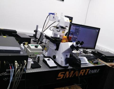
Microscope: Fully motorized and computer-controlled Zeiss Axio-Observer Z1 microscope (inverted) with motorized stage and proprietary Definite Focus technology. Piezo-driven stage for scanning stages with a maximum travel range of 250 µm.
Objectives:
- Plan-Apochromat 10X/0.45 NA (air)
- Plan-Apochromat 20X/0.8 NA (air)
- C-Apochromat 40x/1.2 NA (water) for FCS measurements
- Plan-Apochromat 40x/1.3 NA (oil)
- iPlan-Apochromat 63x/1.4 NA (oil)
- iPlan-Apochromat 100x/1.4 (oil) - DIC imaging is possible with the last three objectives.
Scanning Module: Up to 8 frames/sec scanning speed at 512 x 512 pixels. 32 channels in a proprietary GaAsP spectral detector (8nm per channel) and two PMT fluorescence channels available. One of the PMTs is low-noise for imaging in the red/far red. One transmission detector for DIC imaging is available.
Lasers:
- Ar+ laser (458nm, 488nm, and 514 nm @25 mW)
- DPSS laser (561 nm @20 mW)
- HeNe laser (633 nm @5 mW)
- Titanium Sapphire multiphoton laser (690 nm - 1040 nm with 2.5W @800nm)
Filters: DAPI (only through multiphoton laser), Alexa Fluor 488, Rhodamine.
Zen 2012 acquisition software from Zeiss with 3D, ROI, FRAP, and stitching modules.
Instructions for Registration, Sample Preparation, User Instructions, Precautionary Measures and Charges
Register online through the IRCC webpage.
After the slotbooking request is accepted, please contact the operator (Pradip Shinde or Santosh Panigrahi at 4770) to discuss the details of your experiment.
Currently, we can image fixed samples by sealing them between a glass slide and a cover slip to ensure optimal imaging conditions. It's crucial not to bring samples without this cover slip seal.
For 35 mm diameter petri dishes, it is recommended to use specially designed imaging petri dishes that feature cover slip bottoms, especially when using oil immersion objectives for microscopy.
We shall accept online registration only through the IRCC webpage. If you need to cancel your slot, send an email immediately to with an explanation.
- Slots will be provided on a first-come-first-served basis.
- USB drives are strictly prohibited for copying data to minimize virus-related issues. You are requested to bring a new blank CD to transfer your data. All data must be transferred within 7 days of imaging. Without exception.
- Users must be present during the entire slot.
Applications
Multi-colour imaging, Z-stacks, and 3D image reconstruction are advanced techniques used in microscopy to capture and analyze multiple fluorescent labels in a sample, reconstructing them into three-dimensional models.
Time series imaging involves capturing images at intervals, optionally with Z-stack capabilities, to study dynamic processes over time
Tile scanning automates the imaging of different parts of a sample over extended periods, facilitating large-scale data acquisition and analysis.
Fluorescence techniques like FRET (Fluorescence Resonance Energy Transfer), FRAP (Fluorescence Recovery After Photobleaching), and FCS/FCCS (Fluorescence Correlation Spectroscopy/Fluorescence Cross-Correlation Spectroscopy) are used to study molecular interactions, dynamics, and diffusion in live cells
FLIM (Fluorescence Lifetime Imaging Microscopy) measures the fluorescence decay rates of fluorophores to analyze biochemical processes in biological systems.