External users: registration to be carried out only through I-STEM portal
Additional information about sample and analysis details should be filled in the pdf form provided in the I-STEM portal under “DOWNLOAD CSRF”
Internal users (IITB): registration to be carried out only through DRONA portal
Additional information about sample and analysis details should be filled in the pdf form provided here.
.
Category
- Microscopy and Imaging » Electron Microscopy
Booking Details
TEM Bright field/dark field imaging, HR-TEM imaging,Diffraction pattern
Energy Dispersive spectroscopy (EDS)
Electron Energy Loss spectroscopy (EELS)
Facility Management Team and Location
Dr. Bharati Patro
bharati@iitb.ac.in
Facility Features, Working Principle and Specifications
Features:
Resolution: Point : 2.0 Angstrom
Line : 1.0 Angstrom
Accelerating Voltage: 300 kV
Magnification Range: 58 x to 1 Million x
Accessories
• EDS : EDAX with Octane ELITE T Super
• STEM : on axis BF/DF & HAADF detector
• EELS with GIF (Gatan Image Filter)
Working Principle:
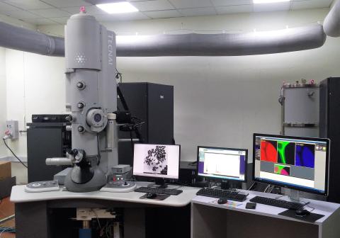
Transmission Electron Microscopy (TEM) is a technique in which a beam of high energy electrons is transmitted through a very thin specimen to form an image which reveals the information about morphology and crystallography. FEG-TEM can go upto atomic scale resolution to view the atomic arrangement of the material.
Scanning Transmission Electron Microscopy (STEM) in which the electron beam is focused to a fine spot to scan in the raster pattern across the specimen and is suitable for analytical techniques such as Z-contrast, annular dark-field imaging, and spectroscopic mapping for chemical composition by Energy Dispersive Spectroscopy (EDS) and as well as Electron Energy Loss Spectroscopy (EELS). These signals can be obtained simultaneously, allowing direct correlation of images and spectroscopic data.
TEM Bright field/dark field imaging, HR-TEM imaging,Diffraction pattern
Energy Dispersive spectroscopy (EDS)
Electron Energy Loss spectroscopy (EELS)
Sample Preparation, User Instructions and Precautionary Measures
- The user has to collect TEM grids from the 300kV FEG TEM lab.
- The user has to come for analysis with a prepared sample on the TEM grid.
- The samples should be prepared on TEM grids of 3mm size and sample thickness should be 50- 100 nm for analysis.
- Any query related to your 300kV FEG-TEM analysis can be emailed to fegtem300@iitb.ac.in.
- The samples should be dry and should withstand ultra-high vacuum
We prefer you or your representative who knows and understands the sample should be present on the day of appointment.
Sample preparation if any should be done at the user end only.
The samples should be dry and should withstand ultra-high vacuum.
Charges for Analytical Services in Different Categories
Field Emission Gun-Transmission Electron Microscope 300kV with STEM, EDS and EELS (FEG-TEM 300kV) charges:
Description | IIT Bombay Users | IITB-Monash Students | Academic Institute / University | National Labs / SINE/IITB Research Park (MSME)(Letter from Research Park required) | IITB Research Park (Big Industries) /Start-up/MSME (Letter from Research Park/Start-up/MSME required) | Industry |
|
No GST | +GST @ 18% | +GST @ 18% | +GST @ 18% | +GST @ 18% | +GST @ 18%
| ||
Imaging (TEM, HR-TEM and Diffraction) | 1500 | 1500 | 3000 | 7500 | 11250 | 15000
| Per Sample
|
EDS analysis | 1200 | 1200 | 2400 | 6000 | 9000 | 12000
| Per EDS |
EDS Mapping | 1200 | 1200 | 2400 | 6000 | 9000 | 12000 | Per Map
|
EDS Line Scan | 1200 | 1200 | 2400 | 6000 | 9000 | 12000 | Per Line Scan
|
EELS analysis | 2250 | 2250 | 4500 | 11250 | 16875 | 22500
| Per ROI |
Applications
- Nano science/Nano Technology
- Micro/Nano electronics
- Thin Films
- Catalysis
- Corrosion
- Polymer science
- Energy science/Engg.
- biological and life sciences
Sample Details
NA
NA
NA
NA
NA
If samples are not dried, column vacuum gets contaminated
Sample thickness should be less than 100 nm for TEM / HR-TEM / Diffraction Imaging and STEM-EDS analysis.
For EELS analysis sample thickness is expected to be around 50 nm
SOP, Lab Policies and Other Details
Publications
- Influence of high concentration of Bi dopant on the structural and optical properties of CsPbBr3 perovskite nanocrystals, Swati Mamgain & Aswani Yella, Emergent Materials (2023 )
- Iron tolerant Bacillus badius mediated bimetallic magnetic iron oxide and gold nanoparticles as Doxorubicin carrier and for hyperthermia treatment, Megha P. Desai , Ana C. Paiva Santos , Mansingraj S. Nimbalkar ,Kailas D. Sonawane , Pramod S. Patil , Kiran D. Pawar, Journal of Drug Delivery and technology, Volume 81,104214 (2023)
- Synthesis and characterization of CdSe decorated ZnO microtubes and their application in dye degradation under visible light, Priya Drimri, Jyoti Rawat, Anubhi Semwal, Himani Sharma, Charu Dwivedi, Materials Today: Proceedings, (2023 )
- Flow synthesis of intrinsically radio labeled and renal-clearable ultrasmall [198Au] Au nanoparticles in a PTFE microchannel, Rubel Chakravarty , Nirvik Sen , Sanchita Ghosh ,Haladhar, Chemical Engineering Journal Advances ,Volume 14, 100456 (2023)
- Antiproliferiative Activity of Biogenic Silver Nanoparticles Synthesized from Leonotis nepetifolia (L) on Human Cancer Cell lines, M. N. L. C. Harika and Parvataneni Radhika,Indian J Pharm Sci; 85(1); 152-163(2023)
- Effect of rolling texture on the precipitation and mechanical behaviour of maraging steels, Kevin Jacob, Heena Khanchandani, Saurabh Dixit, Balila Nagamani Jaya, SSRN, (2023)
- Germanium-Free Dense Lithium Superionic Conductor and Interface Re-Engineering for All-Solid-State Lithium Batteries against High-Voltage Cathode,Govind Kumar Mishra,Manoj Gautam,K. Bhawana,Nilanjan Chakrabarty and Sagar Mitra,ACS Appl. Mater. Interfaces, 15, 8, 10629–10641 (2023)
- Novel hydroxyapatite nanoparticle-based antibiotic alternative to combat methicillin-resistant S. aureus: A mechanism by targeting the structural and functional stability of MRSA membrane protein, Kavita Kadu,Vijay Hemmadi, Malabika Biswas, Meenal Kowshik & Sutapa Roy Ramanan ,Journal of Materials Research (2023 )
- Fabrication of dual functional 3D-flower shaped NaYF4:Dy3+/Eu3+ and graphene oxide based NaYF4:Dy3+/Eu3+ nanocomposite material as a potable luminescence sensor and photocatalyst for environmental pharmaceutical pollutant nitrofurazone in aquatic medium,Richa Singhaal, Nargis Akhter Ashashi, Charanjeet Sen, Swaita Devi, Haq Nawaz Sheikh,Nano-Structures & Nano-Objects,Volume 34, 100965(2023)
- Tuning the mechanical and thermal properties of (MgNiCoCuZn)O by intelligent control of cooling rates,Varatharaja Nallathambi, Lalithkumar Bhaskar, Di Wang, Aleksandr. Naberezhnov, SergeyV. Sumnikov, Emanuel Ionescu, Ravi Kumar, Journal of the European Ceramic Society (2023)
- Photocatalytic hydrogen production, dye degradation, and antimicrobial activity by Ag-Fe co-doped Tio2 nanoparticles, G. K. Sukhadeve, Harshit Bandewar, S.Y. Janbandhu, J.R. Jayaramaiah, R.S. Gedam, Journalof Molecular Liquids, Volume 369, 120948 (2023)
- Sonocatalytic Degradation of Tetracycline with Cu-Doped TiO2 Nanoparticles as the Catalyst: Optimization, Kinetics, and Mechanism, Ansaf V. Karim, Sukanya Krishnan & Amritanshu Shriwastav, Water, Air & Soil Pollution, volume 234, Article number: 41 (2023)
- Ru–W modified graphitic carbon nitride by a monomer complexation synthesis approach from a tailored polyoxometalate: towards electrochemical detection of hydrogen peroxide released by cells,Neermunda Shabana, Ajith Mohan Arjun, K. Rajendran, Soyeb Pathan and P. Abdul Rasheed, Royal society of chemistry, Anal. Methods, 15, 587-595(2023)
- An attempt to enhance the afterglow luminescence of NIR light emitting long persistent phosphor Zn3Ga2Ge2O10:Cr3+ by Pr3+ co-doping, Birendra Kumar Rajwar, Jairam Manam, Shailendra Kumar Sharma, Spectrochimica Acta Part A: Molecular and Biomolecular Spectroscopy, Volume 293, 122512(2023)
- Photocatalytic performance of glasses embedded with Ag-TiO2 quantum dots on photodegradation of indigo carmine and eosin Y dyes in sunlight, S.Y. Janbandhu, Umakanta Patra, G.K. Sukhadeve, Rahul Kumar, R.S.Gedam, Inorganic Chemistry Communications, Volume 148, 110317 (2023)
- Core-shell structured Fe3O4@MgO: magnetically recyclable nanocatalyst for one-pot synthesis of polyhydro-quinoline derivatives under solvent-free conditions, Gayatree Shinde & Jyotsna Thakur, Journal of Chemical Sciences volume 135, Article number: 14 (2023)
- Palladium nanoparticles-confined pore-engineered urethane-linked thiol-functionalized covalent organic frameworks: a high-performance catalyst for the Suzuki Miyaura cross-coupling reaction, Falguni Shukla , Miraj Patel , Qureshi Gulamnabi and Sonal Thakore , Royal society of chemistry, 10.1039/D2DT04057C (Paper) Dalton Trans., 52, 2518-2532 (2023)
- In–situ synthesis of MnO dispersed carbon nanofibers as binder-free electrodes for high-performance super-capacitors, Shriram Radhakanth, Richa Singhal, Chemical Engineering Science, Volume 265, 118224 (2023)
- The smallest anions entrapped mayenite electride@graphitic carbon core-shells reinforced with superparamagnetic Fe3O4 delivers unrivalled high-frequency microwave absorption, Vidhya Lalan, Subodh Ganesanpotti, Chemical Engineering Journal, Volume 461, 141857 (2023)
- Chlorophyll sensitized and salicylic acid functionalized TiO2 nanoparticles as a stable and efficient catalyst for the photocatalytic degradation of ciprofloxacin with visible light, Sukanya Krishnan, Amritanshu Shriwastav, Environmental Research, Volume 216, Part 2, 114568 (2023)
- Oxygen deficient Ce doped CO supported on alumina catalyst for low-temperature CO oxidation in presence of H2O and SO2,Jyoti Waikar, Pavan More, Fuel, Volume 331, Part 2, 125880 (2023)
- Luminescent and photocatalytic activity of NaGd(MoO4)2: Dy3+/Eu3+ and NaGd(WO4)2: Dy3+/Eu3+ nanorods for efficient sensing and degradation of the antibiotic drug, nitrofurantoin, Swaita Devi , Richa Singhaal , Charanjeet Sen and Haq Nawaz Sheikh, Royal society of chemistry, 10.1039/D2NJ05980K, New J. Chem, 47, 4949-4963 (2023)
- One-step hydrothermal synthesis of WS2 as efficient electrode material for energy storage application, Anuprava Mandal, Nivedita Pandey, Subhananda Chakrabarti, Proc. SPIE 12423, 2D Photonic Materials and Devices VI, 124230E,doi: 10.1117/12.2649635, (2023)
- A facile one step hydrothermal synthesis of flower-like nanosheets of MoS2 for nanoelectronics technology, Anuprava Mandal, Nivedita Pandey, Subhananda Chakrabarti, Proc. SPIE 12423, 2D Photonic Materials and Devices VI, 124230D; doi: 10.1117/12.2648841 (2023)
- Facile hydrothermal synthesis of vanadium disulfide nanomaterial for supercapacitor application, Anuprava Mandal, Nivedita Pandey, Sushil Kumar Pandey, Ashish Kumar Yadav, Subhananda Chakrabarti, Proc. SPIE 12423, 2D Photonic Materials and Devices VI, 124230F doi: 10.1117/12.2649638 (2023)
- Bimetallic CoNi Nanoflowers for Catalytic Transfer Hydrogenation of Terminal Alkynes Neha Choudhary,VireshKumar, and ShaikhM.Mobin,Chemistry Europe, doi.org/10.1002/slct.202202501(2023)
- Iron tolerant Bacillus badius mediated bimetallic magnetic iron oxide and gold nanoparticles as Doxorubicin carrier and for hyperthermia treatment,Megha P. Desai, Ana C. Paiva-Santos, Mansingraj S. Nimbalkar, Kailas D. Sonawane, Pramod S. Patil, Kiran D. Pawar, Journal of Drug Delivery Science and Technology, Volume 81, 104214 (2023)
- Development of a Lemongrass/Silver Nanocomposite for Controlling a Foodborne Pathogen - Escherichia coli Shipra Pandey, Kajal Sharma, and Venkat Gundabala, ACS Food Sci. Technol. 2022, 2, 12, 1850–1861, (2022 )
- Dopant-Free Main Group Elements Supported Covalent Organic− Inorganic Hybrid Conducting Polymer for Sodium-Ion Battery Application Seenuvasan Vedachalam, Pandiaraj Sekar, Chandrasekaran Nithya,Nithya Murugesh, and Ramasamy Karvembu, ACS Appl. Energy Mater. 2022, 5, 1, 557–566 (2022)
- Metal−Dielectric Interfacial Engineering with Mesoporous NanoCarbon Florets for 1000-Fold Fluorescence Enhancements: Smartphone-Enabled Visual Detection of Perindopril Erbumine at a Single-molecular Level Seemesh Bhaskar, Dipin Thacharakkal, Sai Sathish Ramamurthy, and Chandramouli Subramaniam, ACS Sustainable Chem. Eng. 2023, 11, 1, 78–91 (2022)
- Direct-Contact Prelithiation of Si−C Anode Study as a Function of Time, Pressure, Temperature, and the Cell Ideal Time, Manoj Gautam, Govind Kumar Mishra, Aakash Ahuja, Supriya Sau, Mohammad Furquan, and Sagar Mitra, ACS Appl. Mater. Interfaces, 14, 15, 17208–17220, (2022)
- Synthesis and characterization of CdSe decorated ZnO microtubes and their application in dye degradation under visible light, Priya Dimri, Jyoti Rawat, Anubhi Semwal, Himani Sharma, Charu Dwivedi, Materials Today: Proceedings (2023)
- Investigation of microstructure and mechanical properties of microwave consolidated TiMgSr alloy prepared by high energy ball milling, N.B. Pradeep, M.M.Rajath Hegde, Shashanka Rajendrachari, A.O. Surendranathan, Powder Technology, Volume 408, 117715 (2022)
- Antimicrobial activity of synthesized graphene oxide‑selenium nanocomposites: A mechanistic insight Isha Riyal, Ayush Badoni, Shubham S. Kalura,Kavita Mishra,Himani Sharma,Lokesh Gambhir, Charu Dwivedi, Environmental Science and Pollution Research volume 30, pages19269–19277 (2023)
- Nucleotide(s)-mediated simultaneous N, P co-doped reduced graphene oxide (N, P-rGO) porous nanohybrids as high-performance electrode materials for designing sustainable binder-free high-voltage (2.8 V) aqueous symmetric supercapacitors and electrochemical sensors,Ikrar Ahmad and Anil Kumar, Royal society of chemistry (2022)
- Spherical Silver Nanocrystals Arranged in a Metastable Square Pattern Sushil Swaroop Pathak, Savita Priya, Gotluru Kedarnath, and Leela S. Panchakarla, ACS Omega 2022, 7, 32, 28481–28486, (2022)
- Hybrid photoluminescent material from lanthanide fluoride and graphene oxide with strong luminescence intensity as a chemical sensor for mercury ions,Richa Singhaal , Lobzang Tashi, Swaita Devi and Haq Nawaz Sheikh, Royal society of chemistry, New J. Chem., 46, 6528-6538,(2022)
- Effect of layer-by-layer synthesized graphene– polyaniline-based nanocontainers for corrosion protection of mild steel, Siddhant Varshney, Karan Chugh, and S. T. Mhaske, Journal of Materials Science volume 57, pages8348–8366 (2022)
- Mixed-Ligand Assisted Direct Synthesis of Redox-Active UiO-66-(SH)2 Metal Organic Frameworks and Their Behavioural Pattern in Reductive and Oxidative Environments, Sumanta Chowdhury , Parul Sharma , Koustav Kundu , Partha Pratim Das , Preeti Rathi , Prem Felix Siril,Materials Chemistry,(2022)
- Synthesis of silver nanoparticles using underutilized fruit Baccaurea ramiflora (Latka) juice and its biological and cytotoxic efficacy against MCF-7 and MDA-MB 231 cancer cell lines, Swarnendra Banerjee, Shehnaz Islam, Ansuman Chattopadhyay,Arnab Sen, Pallab Kar, South African Journal of Botany Volume 145, Pages 228-235 (2022)
- One-Pot In Situ Synthesis of Mn3O4/N-rGO Nanohybrids for the Fabrication of High Cell Voltage Aqueous Symmetric Supercapacitors: An Analysis of Redox Activity of Mn3O4 toward Stabilizing the High Potential Window in Salt-in-Water and Water-in-Salt Electrolytes,Sahil Thareja and Anil Kumar, nergy Fuels 2022, 36, 24, 15177–15187,Publication (2022)
- Continuous flow scale-up of biofunctionalized defective ZnO quantum dots: A safer inorganic ingredient for skin UV protection, Sayoni Sarkar, SujitKumar Debnath, Rohit Srivastava, Ajit R. Kulkarni, Acta Biomaterialia, Volume 147, Pages 377-390 (2022)
- The biosynthesis of nickel oxide nanoparticles using watermelon rind extract and their biophysical effects on the germination of Vigna radiata seeds at various concentrations, Mohd Kashif Aziz, Sudhakar Chauhan, Zeba Azim, Gyanendra Kumar Bharati and Shekhar Srivastava, International Journal of Science and Research Archive, 07(02), 245–254 (2022)
- Sono‑assisted synthesis of AgFeO2 nanoparticles for efcient removal of Basic Green‑4 dye from aqueous solution, Maheshwari Zirpe, Jyotsna Thakur,Journalof Nanoparticle Research volume 24, Article number: 240 (2022)
- Polypyrrole and a polypyrrole/nickel oxide composite – single-walled carbon nanotube enhanced photocatalytic activity under visible light,Prasenjit Chakraborty , Sk. Taheruddin Ahamed , Pinaki Mandal , Anup Mondal and Dipali Banerjee, Royal society of chemistry, New J. Chem., 46, 14065-14080 (2022)
- The synergetic effect of PdCr based bimetallic catalysts supported on RGO-TiO2 for organic transformations, Nitika Sharma, Chandan Sharma, Shally Sharma, Sukanya Sharma, Satya Paul, Results in Chemistry, Volume 4, 100524, (2022)
- Influence of solvents, reaction temperature, and aging time on the morphology of iron oxide nano particles, Prashil K. Narnaware & C. Ravikumar, INORGANIC AND NANO-METAL CHEMISTRY2022, VOL. 52, NO. 7, 922–936, (2022)
- The smallest anions, induced porosity and graphene interfaces in C12A7:e− electrides: a paradigm shift in electromagnetic absorbers and shielding materials,Vidhya Lalan and Subodh Ganesanpotti , J. Mater. Chem. C, 10, 969-982(2022)
- Binder free electrodeposition fabrication of NiCo2O4 electrode with improved electrochemical behavior for supercapacitor application,Manpreet Kaur , Prakash Chand, Hardeep Anand, Journal of Energy Storage, Volume 52, Part B, 104941 (2022)
- Insight into the Modulation of Carbon-Dot Optical Sensing Attributes through a Reduction Pathway,Nirmiti Mate,Pranav,Kallayi Nabeela,Navpreet Kaur,Shaikh M. Mobin, ACS Omega 2022, 7, 48, 43759–43769,Publication (2022)
- Magnetically Recyclable Ag@Fe2O3 Core-shell Nanostructured Catalyst for One-pot Synthesis of 2-Aryl Benzimidazole and Benzothiazole, Gayatree Shinde and Jyotsna Thakur, Current Organocatalysis, Volume 9, pp. 237-251(15), (2022)
- Energy storage and photosensitivity of in-situ formed silver-copper (Ag-Cu) heterogeneous nanoparticles generated using multi-tool micro electro discharge machining process,Ishwar Bhiradi, Somashekhar S. Hiremath, Journal of Alloys and Compounds,Volume 897, 162950, (2022)
- Bioactive Molecules Coated Silver Oxide Nanoparticle Synthesis from Curcuma zanthorrhiza and HR-LCMS Monitored Validation of Its Photocatalytic Potency Towards Malachite Green Degradation, K. S. Aiswariya & Vimala Jose, Journal of Cluster Science volume 33, pages1685–1696, (2022)
- Mechanistic Pathway of Lipid Phase-Dependent Lipid Corona Formation on Phenylalanine-Functionalized Gold Nanoparticles: A Combined Experimental and Molecular Dynamics Simulation Study,Avijit Maity,Soumya Kanti De,Debanjan Bagchi,Hwankyu Lee,Anjan Chakraborty, J. Phys. Chem. B , 126, 11, 2241–2255 (2022)
- Complete oxidation of CO and propene as model component of diesel exhaust and VOC using manganese oxide supported on octahedral (AlO63−)-Ce3+ ,Waikar, Pavan More, Applied Surface Science Advances, Volume 7, 100204 ,(2022)
- Investigating the oxidative reactivity and nanostructural characteristics of diffusion flame generated soot using methyl crotonate and methyl butyrate blended diesel fuels, Samantha DaCosta , Akshay Salkar , Anand Krishnasamy , Ravi Fernandes , Pranay Morajkar, Fuel, Volu me 309, 122141, (2022)
- Catalytic dye degradation by novel phytofabricated silver/zinc oxide composites, Khalida Bloch , Shahansha M. Mohammed , Srikanta Karmakar , Satyajit Shukla , Adersh Asok, Kaushik Banerjee , Reshma Patil-Sawant, Noor Haida Mohd Kaus , Sirikanjana Thongmee and Sougata Ghosh, Front. Chem. 10:1013077 (2022)
- In situ interfacial nanoengineering of imidazole-bridged one dimensional AgVO3 nanoribbons by Ag fractals, K.K. Sarigamala , T. Albrecht , S. Shukla , S. Saxena, Materials Today Chemistry, (2022)
- Norbornane derived N-doped sp2 carbon framework as an efficient electrocatalyst for oxygen reduction reaction and hydrogen evolution reaction, Rupali S. Mane , Shakeelur Rahema A.R., Tejes Kothawade , Himanshu Chakraborty, Neetu Jha, Fuel 323, 124420 ,(2022)
