External users: registration to be carried out only through I-STEM portal
Additional information about sample and analysis details should be filled in the pdf form provided in the I-STEM portal under “DOWNLOAD CSRF”
Internal users (IITB): registration to be carried out only through DRONA portal
Additional information about sample and analysis details should be filled in the pdf form provided here.
.
Category
- Microscopy and Imaging » Electron Microscopy
Booking Details
Facility Management Team and Location
Facility Features, Working Principle and Specifications
Facility Description
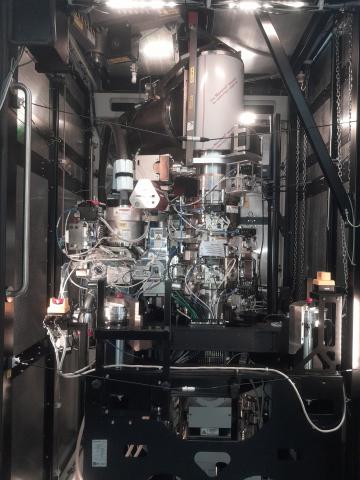
Our facility features a 300 kV Thermo Fisher Krios G4 cryo-electron microscope for high-resolution cryo-EM analysis, equipped with a BioContinuum GATAN K3 detector and an Amtek-Gatan energy filter. This setup supports both single-particle analysis and tomography, with all necessary accessories for preparing frozen-hydrated samples. Additionally, we have a TalosL120C screening microscope and sample preparation tools such as the Vitrobot, Cryo-Ultramicrotome, and High-Pressure Freezer, ensuring efficient sample handling. This advanced infrastructure enables detailed imaging of biomolecules like proteins, viruses, and nanoparticles, providing valuable insights into cellular structures.
Cryo-Electron Microscopy (Cryo-EM) is a technique used to capture high-resolution 3D images of biological samples in their frozen, near-native state. The process begins by rapidly freezing the sample in vitreous ice to preserve its structure. The sample is then imaged using an electron microscope, which captures 2D projection images from multiple angles. These images are computationally processed to generate a 3D reconstruction of the sample. Cryo-EM enables the visualization of macromolecules at near-atomic resolution, making it ideal for studying complex biological structures like proteins, protein complexes, and viruses. This method eliminates the need for crystallisation or staining, providing detailed insights into molecular architecture.
Microscope: Titan Krios G4
Imaging: Utilizing an Extreme High-Brightness Field Emission Gun (X-FEG) and offering a flexible accelerating voltage range from 80 to 300 kV, complete with automatic condenser, objective, and SA apertures to facilitate multiple image acquisitions per grid hole.
Detector: Equipped with both BioContinuum GATAN K3 detector and Ceta D detectors.
Energy Filter: Incorporating the Biocontinuum K3 energy filter.
Software for Data Collection and Screening: Utilizing EPU for streamlined data collection and screening processes.
Sample Preparation, User Instructions and Precautionary Measures
Before booking a slot for the Krios 300kV microscope, users must first examine the sample on a negatively stained grid, followed by screening on a cryo-conditioned grid using the Talos 120kV microscope. The corresponding data should be submitted when reserving the slot.
- Users are required to complete online registration.
- External users must pre-register.
- Appointments will be scheduled according to the queue and confirmed via email.
- New users should contact the CryoEM lab before registering.
- Users must notify the CryoEM lab at least 24 hours in advance if they wish to cancel or reschedule their appointment.
Charges for Analytical Services in Different Categories
Applications
- Structural Biology: It helps visualize protein complexes, viruses, and macromolecular interactions at near-atomic resolution.
- Drug Discovery: Cryo-EM aids in identifying drug targets and designing therapeutics by revealing detailed biomolecular structures.
- Cell Biology: It allows the study of cellular architecture and dynamics through cryo-tomography, including organelles and protein interactions.
- Virology: Cryo-EM is essential for studying viral structure and interactions with host cells, contributing to vaccine and drug development.
- Nanotechnology: It is used to analyze nanoparticles and nanomaterials for biomedical and technological applications.
- Neuroscience: Cryo-EM helps investigate synaptic structures and neurodegenerative diseases.
- Microbial Research: It provides insights into bacterial structures and protein folding, aiding the study of antibiotic resistance and disease mechanisms.
- Materials Science: Cryo-EM is employed to study the atomic structures of materials and nanostructures.
- Cryo-Tomography: It enables 3D imaging of cells and tissues, preserving their native state.
- Functional Studies: Cryo-EM captures conformational changes in proteins, aiding the understanding of their biological functions.
