External users: registration to be carried out only through I-STEM portal
Additional information about sample and analysis details should be filled in the pdf form provided in the I-STEM portal under “DOWNLOAD CSRF”
Internal users (IITB): registration to be carried out only through DRONA portal
Additional information about sample and analysis details should be filled in the pdf form provided here.
.
Category
- Microscopy and Imaging » Electron Microscopy
Booking Details
EDS-Elemental mapping
STEM-Brightfield and darkfield images Electron diffraction for crystallography studies
Facility Management Team and Location
Facility Features, Working Principle and Specifications
Facility Description
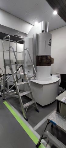
This facility facilitates TEM analysis of biological as well as soft solids on cryo mode or Room temperature mode. This facility works on Cryo EM technique where biological samples are preserved in vitreous ice and imaged by EM at cryogenic temperatures. High contrast helps in imaging biological samples without staining.
Working Principle
Aberration corrected Cryo-HRTEM
Make and Model: JEOL NEOARM 200F
"NEOARM" / JEM-ARM200F comes with unique cold field emission gun (Cold FEG) and a new Cs corrector (ASCOR) that compensates for higher order aberrations. The combination of a Cold FEG and ASCOR enables atomic-resolution imaging at not only 200 kV accelerating voltage, but also a low voltage of 30 kV.
"NEOARM" is also equipped with an automated aberration correction system that incorporates aberration correction algorithm for automatic fast and precise aberration correction. This system enables higher-throughput atomic-resolution imaging.
Furthermore, a new STEM detector that provides enhanced contrast of light elements is incorporated as a standard unit. Contrast enhancement of light elements is achieved by a new STEM imaging technique, facilitating observation of light-element materials, even at low accelerating voltages.Cryo-HRTEM
The JEM-2100 is a multipurpose, 200 kV analytical electron microscope with LaB6 source. supports research and development in wide scientific fields, for biology to materials researches.
Cryo-Ultramicrotome
Make and Model: Leica EM UC7
Useful to prepare high-quality ultra- or semi-thin sections for your transmission electron or light microscope investigation whilst simultaneously creating perfectly smooth block face surfaces for atomic force, scanning electron, or incident light microscopy. For ultrathin cryo- sections or surfacing of cryogenic material, EM UC7 ultramicrotome can be equipped with the EM FC7 low-temperature sectioning system as needed.
4. Freeze fracture
Make and Model: Leica EM ACE900
Freeze fracture describes the technique of breaking a frozen specimen to reveal internal structures. Freeze etching is the sublimation of surface ice under vacuum to reveal details of the fractured face that were originally hidden. A metal/carbon mix enables the sample to be imaged in a SEM (block-face) or TEM (replica).
5. Cryo-plunger
Make and Model: Leica EM GP2
System offers automated vitrification to provide fast, easy, and reproducible sample preparation for cryo-EM. It performs the cryo-fixation process at constant physical and mechanical conditions like temperature, relative humidity, blotting conditions, and freezing velocity. This ensures high-quality cryo-fixation results and a high sample preparation throughput prior to cryo-TEM observation.
Transmission Electron Microscope Configuration:
- Point resolution: 0.27 nm
- Lattice resolution: 0.14 nm
- STEM resolution: 0.082 nm
- Electron Source: Cold field emission gun.
- Accelerating Voltage: 80 kV & 200 kV
- Magnification: X2000 - X1500000
- Specimen Tilt angle X/Y: +- 35°
Sample Preparation, User Instructions and Precautionary Measures
Charges for Analytical Services in Different Categories
Description | IITB User | IITB Monash | Academic Institutes | National Lab / R&Ds; SINE | IITB Research Park (Big Industries) /Start-up/MSME (Letter from Research Park/Start-up/MSME required) | Industry | Unit |
GST | Not applicable | 18% | 18% | 18% | 18% | 18% | — |
Imaging (Cryo/RT) / STEM / Tomography / EDS Analysis | ₹1000 | ₹1000 | ₹2000 | ₹5000 | ₹7500 | ₹10000 | Per hour |
Applications
Nanotechnology, Biology and Life sciences, Material science, Pharmaceutical analysis, Semiconductors.
Sample Details
SOP, Lab Policies and Other Details
Publications
List of publications
1. In Vivo Analysis of Biodegradable Liposome Gold Nanoparticles as Efficient Agents for Photothermal Therapy of Cancer AK Rengan, AB Bukhari, A Pradhan, R Malhotra, R Banerjee, Nano letters 15 (2), 842-848
2. Mayur K Temgire, Akkihebbal K. Suresh, Shantaram G. Kane,and Jayesh Bellare Establishing The Interfacial Nano-structure And Elemental Composition of Homeopathic Medicines Based on Inorganic Salts: A Scientific Approach - September 2015 10.1016/j.homp.2015.09.006
3. Formation of nanoparticles and nanocrystals of mercury by α-lactalbumin Chebrola Pulla Rao. Atul Gajanan Thawari . Vijaya Kumar hinge · Mayur Temgire RSC Advances 09/2014; DOI:10.1039/C4RA07156E
4. Chain length dependence of polyol synthesis of zinc ferrite nanoparticles: Why is diethylene glycol so different? Supriya N Rishikeshi· Satyawati S Joshi · Mayur K Temgire · Jayesh R Bellare, Dalton Transactions 02/2013; 42(15). DOI:10.1039/c2dt32026f
5. Redox Decomposition of Silver Citrate Complex in Nanoscale Confinement: An Unusual Mechanism of Formation and Growth of Silver Nanoparticles Sabyasachi Patra†, Ashok K. Pandey†, Debasis Sen‡, Shobha V. Ramagiri§, Jayesh R. Bellare§, S. Mazumder‡, and A. Goswami*† langmuir, 2014, 30 (9), pp 2460–2469
6. Enhancing cubosome functionality by coating with a single layer of poly-ε-lysine Deshpande S1, Venugopal E, Ramagiri SV, Bellare JR, Kumaraswamy G, Singh N.Volume 6, Issue 19, 8 October 2014, Pages 17126-17133 ACS Appl. Mater. Interfaces
7. Aravind Kumar Rengan, Amirali B Bukhari, Arpan Pradhan, Renu Malhotra, Rinti Banerjee, Rohit Srivastava, Abhijit De, (2015) Invivo analysis of biodegradable liposomes gold nanoparticles as efficient agents for photothermal therapy of cancer. Nanoletters, 12, 19.
8. Supriya N Rishikeshi, Satyawati S Joshi, Mayur K Temgire, Jayesh R Bellare, Chain length dependence of polyol synthesis of zinc ferrite nanoparticles: why is diethylene glycol so different? Dalton Trans 2013 Apr 19;42(15):5430-8.
9. Chhabra, H., Gupta, P., Verma, P.J., Jadhav, S., Bellare, J.R.;Gelatin-PMVE/MA composite scaffold promotes expansion of embryonic stem cells; (2014) Materials Science and Engineering C, 37 (1), pp. 184-194.
10. Jaiswal, A.K., Dhumal, R.V., Ghosh, S., Chaudhari, P., Nemani, H., Soni, V.P., Vanage, G.R., Bellare, J.R.; Bone healing evaluation of nanofibrous composite scaffolds in rat calvarial defects: A comparative study; (2013) Journal of Biomedical Nanotechnology, 9 (12), pp. 2073-2085.
11. Jaiswal, A.K., Dhumal, R.V., Bellare, J.R., Vanage, G.R.; In vivo biocompatibility evaluation of electrospun composite scaffolds by subcutaneous implantation in rat; (2013) Drug Delivery and Translational Research, 3 (6), pp. 504-517.
12. Singh, R., Wagh, P., Wadhwani, S., Gaidhani, S., Kumbhar, A., Bellare, J., Chopade, B.A.; Synthesis, optimization, and characterization of silver nanoparticles from Acinetobacter calcoaceticus and their enhanced antibacterial activity when combined with antibiotics; (2013) International Journal of Nanomedicine, 8, pp. 4277-4290.
13. Sagar, N., Pandey, A.K., Gurbani, D., Khan, K., Singh, D., Chaudhari, B.P., Soni, V.P., Chattopadhyay, N., Dhawan, A., Bellare, J.R.; In-Vivo Efficacy of Compliant 3D Nano-Composite in Critical-Size Bone Defect Repair: A Six Month Preclinical Study in Rabbit; (2013) PLoS ONE, 8 (10), art. no. e77578,
14. Kanitkar, M., Jaiswal, A., Deshpande, R., Bellare, J., Kale, V.P.; Enhanced Growth of Endothelial Precursor Cells on PCG-Matrix Facilitates Accelerated, Fibrosis-Free, Wound Healing: A Diabetic Mouse Model; (2013) PLoS ONE, 8 (7), art. no. e69960. </li>
2019-2020
1. Effects of Ethanol Addition on the Size Distribution of Liposome Suspensions in Water, Ankush Pal, P. Sunthar, D. V. Khakhar*, Ind. Eng. Chem. Res. 2019, 58, 18, 7511–7519
2. Efficient separation of biological macromolecular proteins by polyethersulfone hollow fiber ultrafiltration membranes modified with Fe3O4 nanoparticles …, A Modi, J Bellare - International journal of biological macromolecules, 2019 - Elsevier
3. The consequence of silicon additive in isothermal decomposition of hydrides LiH, NaH, CaH2 and TiH2 R Kalamkar, V Yakkundi, A Gangal - … Journal of Hydrogen Energy, 2020
4. Synthesis and properties of amino and thiol functionalized graphene oxide, HP Manwatkar, SD Gedam, CS Bhaskar… - Materials Today …, 2020 - Elsevier
5. Impact of thermal annealing inducing oxidation process on the crystalline powder of In 2 S 3, A Timoumi, W Zayoud, A Sharma, M Kraini… - Journal of Materials …, 2020 - Springer
6. Unique Structure-Induced Magnetic and Electrochemical Activity in Nanostructured Transition Metal Tellurates Co1 – xNixTeO4 (x = 0, 0.5, and 1)copyright © 2020 American Chemical Society
7. Amoxicillin removal using polyethersulfone hollow fiber membranes blended with ZIF-L nanoflakes and cGO nanosheets: Improved flux and fouling-resistance, A Modi, J Bellare - Journal of Environmental Chemical Engineering, 2020 - Elsevier
8. La/Ce mixed metal oxide supported MWCNTs as a heterogeneous catalytic system for the synthesis of chromeno pyran derivatives and assessment of green …, RA Rather, S Siddiqui, WA Khan, ZN Siddiqui - Molecular Catalysis, 2020 - Elsevier
9. Compositional Control as the Key for Achieving Highly Efficient OER Electrocatalysis with Cobalt Phosphates Decorated Nanocarbon Florets, J Saha, S Verma, R Ball, C Subramaniam… - Small, 2020 - Wiley Online Library
10. Hybrid silver–gold nanoparticles suppress drug resistant polymicrobial biofilm formation and intracellular infection, E Bhatia, R Banerjee - Journal of Materials Chemistry B, 2020 - pubs.rsc.org
11. Development of oxidation resistant and mechanically robust carbon nanotube reinforced ceramic composites, V Verma, SC Galaveen, L Gurnani… - Ceramics …, 2020 - Elsevier
12. Zeolitic imidazolate framework-67/carboxylated graphene oxide nanosheets incorporated polyethersulfone hollow fiber membranes for removal of toxic heavy …, A Modi, J Bellare - Separation and Purification Technology, 2020 - Elsevier
13. Enhanced wettability and photocatalytic activity of seed layer assisted one dimensional ZnO nanorods synthesized by hydrothermal method, D Upadhaya, DD Purkayastha - Ceramics International, 2020 - Elsevier
14. Di-tert-butylphosphate Derived Thermolabile Calcium Organophosphates: Precursors for Ca(H2PO4)2, Ca(HPO4), α-/β-Ca(PO3)2, and Nanocrystalline Ca10(PO4)6 …, S Verma, R Murugavel - Inorganic Chemistry, 2020 - ACS Publications
For the Year 2021
1. Probing Kinetics and Mechanism of Formation of Mixed Metallic Nanoparticles in a Polymer Membrane by Galvanic Replacement between Two Immiscible Metals ... NG Gaidhani, S Patra, HS Chandwadkar, D Sen... - Langmuir, 2021 - ACS Publication. ...
2. Influence of carbon nanotube type and novel modification on dispersion, melt‐rheology and electrical conductivity of polypropylene/carbon nanotube composites J Banerjee, S Kummara, AS Panwar... - Polymer ..., 2021 - Wiley Online Library
3. Composition uniformity and large degree of strain relaxation in MBE-grown thick GeSn epitaxial layers, containing 16% Sn J Rathore, A Nanwani, S Mukherjee... - Journal of Physics D ..., 2021 - iopscience.iop.org
4. Hydrothermal-assisted synthesis and stability of multifunctional MXene nanobipyramids: structural, chemical, and optical evolution B Singh, R Bahadur, S Neekhra, M Gandhi... - ... Applied Materials & ..., 2021 - ACS Publications
5. Influence of carbon nanotube type and novel modification on dispersion, melt‐rheology and electrical conductivity of polypropylene/carbon nanotube composites J Banerjee, S Kummara, AS Panwar... - Polymer ..., 2021 - Wiley Online Library
6. Interaction of Aromatic Amino Acid-Functionalized Gold Nanoparticles with Lipid Bilayers: Insight into the Emergence of Novel Lipid Corona Formation A Maity, SK De, A Chakraborty - The Journal of Physical Chemistry ..., 2021 - ACS Publications
7. Ultra-fast, chemical-free, mass production of high quality exfoliated graphene A Islam, B Mukherjee, KK Pandey, AK Keshri - ACS nano, 2021 - ACS Publications
8. Multifunctional hybrid soret nanoarchitectures for mobile phone-based picomolar Cu2+ ion sensing and dye degradation applications S Bhaskar, P Jha, C Subramaniam... - Physica E: Low ..., 2021 - Elsevier
9. Polyphenol stabilized copper nanoparticle formulations for rapid disinfection of bacteria and virus on diverse surfaces K Sadani, P Nag, L Pisharody, XY Thian, G Bajaj... - ..., 2021 - iopscience.iop.org
10. Bicomponent Coassembled Hydrogel as a Template for Selective Enzymatic Generation of DOPAS Biswas, T Ghosh, DKK Kori, AK Das - Langmuir, 2021 - ACS Publications
11. Improved non-enzymatic H2O2 sensors using highly electroactive cobalt hexacyanoferrate nanostructures prepared through EDTA chelation route R Banavath, R Srivastava, P Bhargava - Materials Chemistry and Physics, 2021 - Elsevier
12. Enhanced antibacterial activity of decahedral silver nanoparticles S Bharti, S Mukherji, S Mukherji - Journal of Nanoparticle Research, 2021 - Springer
13. Non-Enzymatic H2O2 Sensor Using Liquid Phase High-Pressure Exfoliated Graphene R Banavath, SS Nemala, R Srivastava... - Journal of The ..., 2021 - iopscience.iop.org
14. Spontaneous Ion Migration via Mechanochemical Ultrasonication in Mixed Halide Perovskite Phase Formation:
Experimental and Theoretical Insights M Roy, Vikram, Bhawna, U Dedhia... - The Journal of ..., 2021 - ACS Publications
15. Zeolitic imidazolate framework-8 nanoparticles coated composite hollow fiber membranes for CO2/CH4 separation K Sainath, P Kumari, J Bellare - Journal of Environmental Chemical ..., 2021 - Elsevier
16. Non-stoichiometry induced exsolution of metal oxide nanoparticles via formation of wavy surfaces and their enhanced electrocatalytic activity: case of misfit calcium ... KS Roy, C Subramaniam... - ACS Applied Materials & ..., 2021 - ACS Publications
17. Ion-Induced Bending with Applications for High-Resolution Electron Imaging of Nanometer-Sized Samples S Zhang, V Garg, G Gervinskas... - ACS Applied Nano ..., 2021 - ACS Publications
18. Synthesis, Characterization, and Hydrogen Gas Sensing of ZnO/g-C3N4 Nanocomposite A Ibrahim, UB Memon, SP Duttagupta... - Engineering ..., 2021 - mdpi.com
19. Underlying mechanisms for the modulation of self-assembly and the intrinsic fluorescent properties of amino acid- functionalized gold nanoparticles SK De, A Maity, A Chakraborty - Langmuir, 2021 - ACS Publications
20. [HTML] Novel combination of bioactive agents in bilayered dermal patches provides superior wound healing MM Pillai, H Dandia, R Checker, S Rokade... - ... , Biology and Medicine, 2022 - Elsevier... Murine
21. Probing Kinetics and Mechanism of Formation of Mixed Metallic Nanoparticles in a Polymer Membrane by Galvanic Replacement between Two Immiscible Metals ... ..., D Sen, C Majumder, SV Ramagiri, JR Bellare - Langmuir, 2021 - ACS Publications
22. Concomitant Effect of Quercetin-and Magnesium-Doped Calcium Silicate on the Osteogenic and Antibacterial Activity of Scaffolds for Bone Regeneration AM Preethi, JR Bellare - Antibiotics, 2021 - mdpi.com
23. “Viscotaxis”-directed migration of mesenchymal stem cells in response to loss modulus gradient..., V Kumar, D Shah, S Beri, S Das, J Bellare... - Acta biomaterialia, 2021 - Elsevier
24. Matrix dependent spatial distributions of in situ formed rhodium nanostructures in ion-exchange membranes..., AK Pandey, SV Ramagiri, JR Bellare - Materials Today Chemistry, 2021 - Elsevier
25. pH-driven enhancement of anti-tubercular drug loading on iron oxide nanoparticles for drug delivery in macrophages KB Cotta, S Mehra... - Beilstein journal of ..., 2021 - beilstein-journals.org
26. A large area flexible p-type transparent conducting CuS ultrathin films generated at liquid-liquid interface SS Pathak, LS Panchakarla - Applied Materials Today, 2021 - Elsevier
