External users: registration to be carried out only through I-STEM portal
Additional information about sample and analysis details should be filled in the pdf form provided in the I-STEM portal under “DOWNLOAD CSRF”
Internal users (IITB): registration to be carried out only through DRONA portal
Additional information about sample and analysis details should be filled in the pdf form provided here.
.
Category
- Microscopy and Imaging » Force Microscopy
Booking Details
Facility Management Team and Location
Facility Features, Working Principle and Specifications
Facility Description
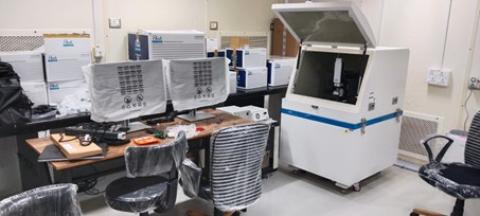
The Atomic Force Microscopy (AFM) Facility is a specialized laboratory equipped for high-resolution surface characterization, nanomechanical measurements, and molecular interactions at the nanoscale. The facility supports a broad range of research applications in materials science, biology, and biophysics.
- Key Features and Capabilities:
- High-Resolution Imaging: Enables topographical mapping of surfaces with nanometer-scale resolution.
- Force Spectroscopy: Measures mechanical properties such as stiffness, elasticity, and adhesion forces at the molecular level.
- Modes of Operation: Includes contact mode, tapping mode, and non-contact mode for diverse sample analysis.
- Environmental Control: Options for imaging in ambient air, liquid environments, and controlled atmospheres for biological and material studies.
- Advanced Data Analysis: Software for quantitative analysis of surface roughness, particle size, and force-distance curves.
- Multi-Sample Compatibility: Capable of analyzing biological samples (cells, membranes, biomolecules), polymers, thin films, and nanomaterials
Atomic Force Microscopy (AFM) operates by scanning a sharp probe (cantilever tip) across a sample surface to measure interactions at the nanoscale. The fundamental principle relies on the detection of forces between the probe and the sample.
- Cantilever & Tip Interaction
- A sharp tip is mounted on a flexible cantilever.
- As the tip approaches the sample, intermolecular forces (van der Waals, electrostatic, etc.) cause the cantilever to deflect.
- Laser-Based Detection System
- A laser beam is focused on the back of the cantilever and reflected onto a position-sensitive photodetector (PSPD).
- Changes in cantilever deflection are recorded to generate surface topography and force measurements.
- Feedback Mechanism
- A feedback loop maintains a constant force or distance between the tip and the sample, ensuring precise imaging.
- Different Imaging Modes
- Non-Contact Mode
- Contact Mode
- Magnetic Force Microscopy (MFM)
- Conductive AFM
- Liquid AFM
- Piezoresponse force microscopy (PFM)
The Atomic Force Microscope (AFM) consists of a rigid metal or composite frame designed for stability and vibration isolation. It features a piezoelectric scanner that enables precise movement, typically allowing an XY scan range of 10–80 µm and a Z range of a few nanometers to micrometers. The cantilever system, with a sharp probe tip, interacts with the sample to capture topographical and mechanical properties. A laser-based detection system measures cantilever deflection, with feedback control ensuring accurate imaging. The AFM also includes an environmental chamber for imaging in air, liquid, or controlled atmospheres, along with advanced data acquisition software for real-time analysis.
Sample Preparation, User Instructions and Precautionary Measures
AFM Sample Preparation Instructions
- Clean the Sample Surface: Ensure the sample is free from dust, debris, and contaminants. Use compressed air, deionized water, or appropriate solvents (e.g., ethanol, acetone) to clean the surface, depending on the sample type.
- Adjust Sample Thickness: The sample should be thin and flat to ensure proper tip approach. If necessary, polish or section thick samples to obtain a smooth and even surface.
- Dry or Hydrate as Needed: For dry samples, ensure complete drying to prevent drift. For liquid-phase imaging (e.g., biological samples), place the sample in an appropriate liquid cell with buffer solution to maintain native conditions.
- Check Sample Compatibility: Ensure the sample is compatible with the selected AFM mode (contact, tapping, or non-contact) and does not degrade under scanning conditions.
- Final Inspection: Before imaging, inspect the sample under an optical microscope to confirm cleanliness and proper adhesion to the holder. Avoid large surface irregularities that could interfere with AFM scanning.
- The experimental data provided is only for research / development purposes. These cannot be used as certificates in legal disputes.
- A maximum 3 samples will be analyzed against a single Registration Form.
- The users should know the approximate roughness or elasticity (For force mode) of the sample before submitting it for analysis.
- MSDS (Material Safety Data Sheet) should be given along with samples to ensure that the samples are not toxic or hazardous. Samples will not be accepted unless accompanied by MSDS.
- Dry sample will be mounted on glass slide with the help of double sided adhesive tape for strong adhesion, Liquid sample will be mounted on 60mm petri dish, sample may get damaged during removal.
- Please add GST at prevailing rates to charges.
Charges for Analytical Services in Different Categories
i. Internal Users: INR 1000 (per 3 h slot) or INR 500 (per 3 h slot) with own AFM tips.
ii. External users
• If the recipient of the report is from Maharashtra: 9% SGST & 9% CGST.
• If the recipient of the report is from outside Maharashtra: 18% IGST.
| Description | Academic | National R & D Lab | Industry/Non Govt. Agencies |
| Per 3Hrs slot (Max sample 3/Slot) | 5000/- | 5000/- | 8000/- |
