Facility Name
External users: registration to be carried out only through I-STEM portal
Additional information about sample and analysis details should be filled in the pdf form provided in the I-STEM portal under “DOWNLOAD CSRF”
Internal users (IITB): registration to be carried out only through DRONA portal
Additional information about sample and analysis details should be filled in the pdf form provided here.
.
Category
- Microscopy and Imaging » Confocal Microscopy
- Microscopy and Imaging » X-ray Microscopy
- Microscopy and Imaging » Optical Microscopy
Booking Details
Facility Management Team and Location
Facility Features, Working Principle and Specifications
Facility Description
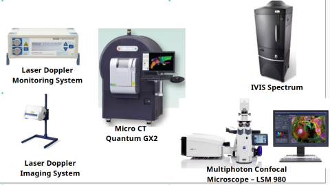
The facility comprise of high end instrument like Micro CT, IVIS, Multiphoton confocal microscope and Laser doppler and monitoring system.
MicroCT (Quantum GX2, Perkin Elmer)
Equipment description: The Quantum GX2 is a basic unit consisting of a Cathode Ray Tube mounted onto a gantry rotor along with detector and emitter, a control and display system, a power supply unit. The main outlet is able to incorporate various bed and bed covers for appropriate sizes of samples. Power supply unit is placed at the back of the system.
Principle: There are cathode ray tubes present in the upper dome of the instrument, whereas below that there is a rotor gantry with detector, emitter, and sample holder, and down in the working mechanics of the equipment. When a particular current with a high voltage hits the cathode tubes, X rays are generated which are focused toward the sample passing through the particular filter. Once the X rays are focused onto the sample, it will absorb, scatter the x ray. The aligned detector is a CMOS camera which will convert the X ray beam into a displayable image onto the screen area. Further this image is analyzed using a particular software.
Applications: Micro-Computed tomography is a useful tool to get sharper images of a sample based on absorption of X-ray for better visualization and representation at a micro level. Micro-CT uses a PMT tube and variety of filter for a wide range of sample type and size. Micro-CT can be used to image and measure bone density and fat contents of the small animals
In vivo Imaging System (IVIS Spectrum, Perkin Elmer)
Equipment description: The IVIS Spectrum comprises a laser, a bed, an anesthesia unit, a set of filters, and a CCD camera detector. Additionally, it includes a PC for display and dedicated software called Living Images for image analysis.
Principle: The IVIS emits laser beams across a range of wavelengths from the UV to far infrared region. When a sample is placed within the field of view (FOV), the laser passes through an excitation filter to obtain desired wavelength light, which interacts with the sample and emits photons. The emitted light passes through an emission filter, allowing the desired wavelength to form an image using the CCD camera.
Applications: IVIS Spectrum facilitates various applications, including:
- Luminescence and fluorescence in vivo sampling
- Bio-distribution studies
- Long-term monitoring of drug effects
Mutiphoton Confocal Microscope (LSM 980, Carl Zeiss)
To analyze samples such as small animals, cells, 3D cell seeded scaffolds etc. with as little disturbance as possible, you must use low labelling density for your biological models. This requires excellent imaging performance combined with low phototoxicity and high speed. LSM 980, your platform for confocal 4D imaging, is optimized for simultaneous spectral detection of multiple weak labels with the highest light efficiency.
Laser Doppler imaging and monitoring system
The moorVMS-LDF laser Doppler monitor for blood flow and temperature monitoring is a high performance, medical grade module for clinic or laboratory. Use of DSP technology brings you a portable, lightweight module featuring uncompromised specification, quality and reliability at a breakthrough price.
The moorLDI2 laser Doppler imager is suitable for a wide range of pre-clinical research investigations, more typically where smaller areas are under investigation. The system features unique focused optics to provide 50 micron pixel size and 512 x 512 pixel resolution for high resolution blood flow images. The scan areas range from just 2.5cm x 2.5cm up to 25cm x 25cm with scan times typically less than 5 minutes. Use of a focussed laser provides a deeper measurement depth, optimal for angiogenesis studies such as hind limb ischemia and tumour modelling and pre-clinical cerebral blood flow imaging. Highly refined image measurement and analysis software allows for flexibility in measurement set up and comprehensive analysis functions. The moorLDI2 features a colour photo image of the scanned area and automatic distance measurement, making the positioning and comparison of images easier.
Sample Preparation, User Instructions and Precautionary Measures
In Vivo Imaging basically uses live animal for imaging hence no prior sample preparation is usually required, however ex vivo imaging protocol require staining which differ according to its applications, which should be discussed briefly with the operator while booking the slot.
Charges for Analytical Services in Different Categories
Charges for Micro CT :
Charges for one hour slot:
| Sr.No. | Category | Micro-CT | IVIS | Multiphoton Microscope | Laser Doppler | ||||
| Inanimate Samples | Live Animals | Inanimate Samples | Live Animals | Inanimate Samples | Live Animals | Tissue blood flow & temperature monitoring | Laser Doppler Imaging System | ||
| 1. | IITB (TAs) | 1000 | 1750 | 500 | 1000 | 500 | 2500 | 1250 | 1500 |
| 2 | IITB Students | 2000 | 3500 | 1000 | 2000 | 1000 | 5000 | 2500 | 3000 |
| 3 | IITB-Monash Students | 2000 | 3500 | 1000 | 2000 | 1000 | 5000 | 2500 | 3000 |
| 4 | Academic Institutes | 4000 | 7000 | 2000 | 4000 | 2000 | 10000 | 5000 | 6000 |
| 5 | National Labs | 10000 | 17500 | 5000 | 10000 | 5000 | 25000 | 12500 | 15000 |
| 6 | Sine (letter from SINE reqd.) | 10000 | 17500 | 5000 | 10000 | 5000 | 25000 | 12500 | 15000 |
| 7 | Research Park (MSME) (letter from RP reqd.) | 10000 | 17500 | 5000 | 10000 | 5000 | 25000 | 12500 | 15000 |
| 8 | Research Park (Big Industry partners) (letter from RP reqd.) and MSME not associated with RP (appropriate certificate required) | 15000 | 26250 | 7500 | 15000 | 7500 | 37500 | 18750 | 22500 |
| 9 | Industries | 20000 | 35000 | 10000 | 20000 | 10000 | 50000 | 25000 | 30000 |
| Note | Per Hour | Per Hour | Per Hour | Per Hour | Per Hour | Per Hour | Per Hour | Per Hour | |
Additional Charges -
- 18% GST on all payments
- For Micro CT, extra hours will be charged as Rs. 1000/- and Rs. 2500/- for inanimate samples and live animal respectively.
- Additional charges for analysis of Micro-CT data will Rs.1000/- sample using Analyze direct software.
- For Multiphoton, extra hours will be charged as Rs. 500/- and Rs. 2500/- for inanimate samples and live animals respectively.
- For Laser Doppler Imaging System, extra hours will be charged as Rs. 2000/-
Important Note:
- For Micro-CT,users should bring contrast agents for imaging, if required.
- For IVIS,user should bring the consumables such as appropriate syringes and dyes for injection.